Journal Information
Vol. 99. Issue 4.
Pages 285-287 (01 October 2023)
Vol. 99. Issue 4.
Pages 285-287 (01 October 2023)
Images in Paediatrics
Full text access
Thymic and pulmonary involvement due to multisystemic Langerhans cell histiocytosis in a 4-month-old boy
Afectación tímica y pulmonar por histiocitosis multisistémica de células de Langerhans en un niño de 4 meses
Visits
882
Boui Meriema,
Corresponding author
Bouimeriemrx@gmail.com

Corresponding author.
, Zouita Btissamb, Lkbir Abidineb, Basraoui Douniab, Jalal Hichamb
a Departamento de Radiología, Hospital Militar Avicena, Marrakech, Morocco
b Departamento de Radiología, Hospital Materno-Infantil de Marrakech, Marrakech, Morocco
This item has received
Article information
Full Text
Bibliography
Download PDF
Statistics
Figures (3)
Show moreShow less
Full Text

Langerhans cell histiocytosis is defined by the proliferation of mononuclear dendritic cells in different tissues or organs1,2; in most cases, it involves bone and skin, while thymic involvement is rare. It usually presents in the paediatric age group. We present the case of a boy with multisystemic Langerhans cell histiocytosis to illustrate thymic and pulmonary involvement in this disease.

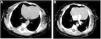
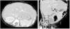
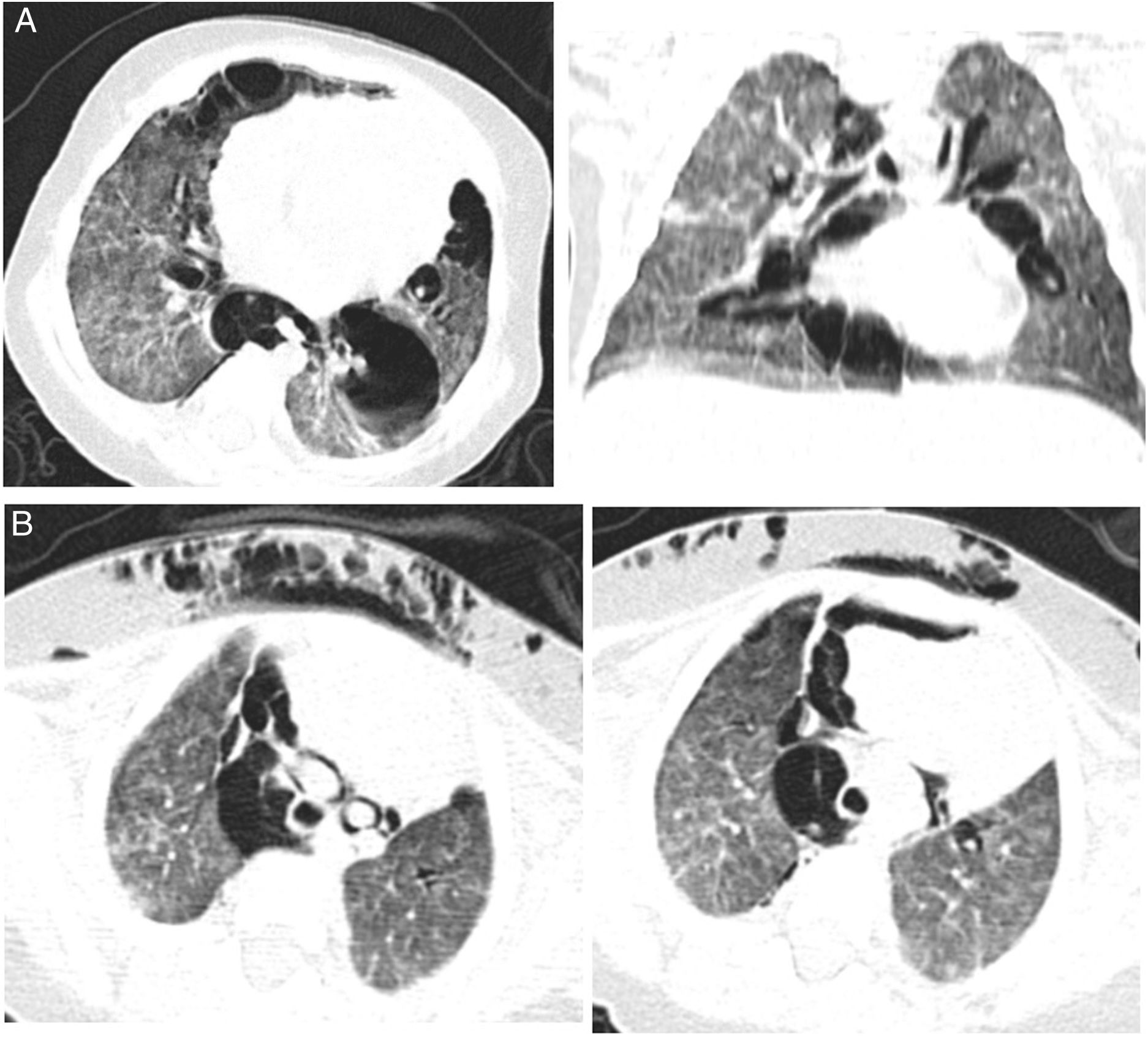
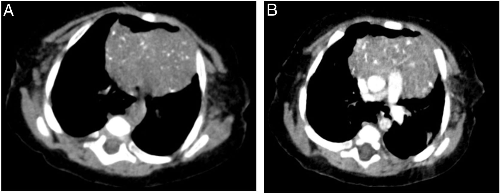
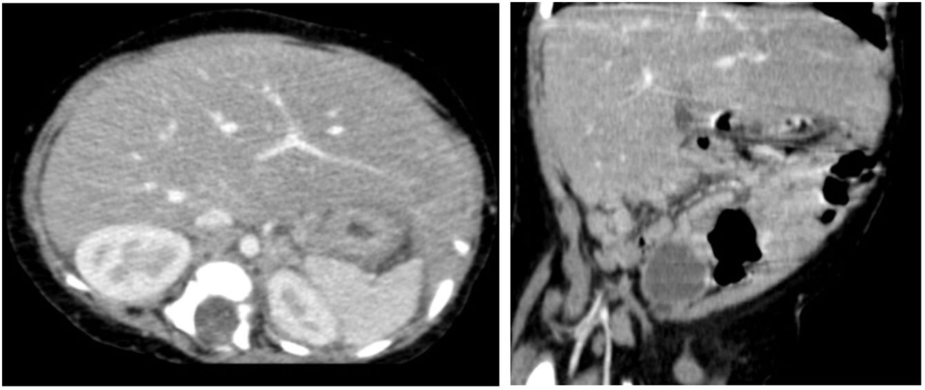
A boy aged 4 months with no history of interest was referred for assessment of generalised cutaneous rashes. Laboratory tests evinced elevation of aminotransferases and alkaline phosphatase. The chest radiograph revealed bilateral diffuse interstitial infiltration. The computed tomography scan of the thorax detected diffuse bilateral interstitial infiltration in the lungs and subpleural thin-walled cysts (Fig. 1A) associated with pneumomediastinum and subcutaneous emphysema (Fig. 1B). The anterior mediastinum was enlarged on account of an enlarged thymus with multiple calcifications (Fig. 2). The computed tomography scan of the abdomen revealed hepatomegaly with a homogeneous pattern (Fig. 3).

Figure 1.

(A) Computed tomography scan in axial and coronal planes showed bilateral diffuse interstitial infiltration in the lungs and subpleural thin-walled cysts. (B) Pneumomediastinum and subcutaneous.

(0.22MB).
Figure 2.

Axial computed tomography scan of the chest without contrast (A) and with contrast (B) evincing thymus hypertrophy. There were multiple punctate calcifications in the thymus, characteristic of Langerhans cell histiocytosis in this organ.

(0.07MB).
Figure 3.

Computed tomography scan of the abdomen in the axial and coronal planes with contrast evinced hepatomegaly with a homogeneous pattern.

(0.07MB).

The computed tomography of the chest and abdomen did not find evidence of regional lymphadenopathy or bone involvement. Langerhans cell histiocytosis was suspected, prompting performance of a skin biopsy, the examination of which revealed Langerhans cell infiltration in the superficial dermis. The specimen was positive for CD1a, langerin and S-100 on immunohistochemistry staining.

The patient received a combination of vinblastine and steroids weekly for 6 consecutive weeks, with a favourable outcome.

Conflicts of interest

The authors have no conflicts of interest to declare.

References
[1]
F.B.E. Favara, F.R. Jaffe.
Pathology of Langerhans cell histiocytosis.
Hematol Oncol Clin North Am, 1 (1987), pp. 75-97
[2]
R. Haupt, M. Minkov, I. Astigarraga, F.E. Schäfer, F.V. Nanduri, F.R. Jubran, et al.
Langerhans cell histiocytosis (LCH): guidelines for diagnosis, clinical work-up, and treatment for patients till the age of 18 years.
Pediatr Blood Cancer., 60 (2013), pp. 175-184
Copyright © 2023. Asociación Española de Pediatría
Idiomas
Anales de Pediatría (English Edition)
Article options
Tools
es en

¿Es usted profesional sanitario apto para prescribir o dispensar medicamentos?

Are you a health professional able to prescribe or dispense drugs?