Journal Information
Vol. 99. Issue 4.
Pages 283-284 (01 October 2023)
Vol. 99. Issue 4.
Pages 283-284 (01 October 2023)
Images in Paediatrics
Full text access
Beetlejuice: acral exogenous pigmentation in a 13-month-old girl
Beetlejuice: pigmentación acral exógena en lactante de 13 meses
Visits
1001
Sonia Milkova Ivanovaa,
Corresponding author
sonia.milkova@salud.madrid.org

Corresponding author.
, Carlos Daniel Grasa Lozanob
a Department of Paediatrics, Hospital Universitario La Paz, Madrid, Spain
b Department of General Paediatrics and Paediatric Infectious Disease, Hospital Universitario La Paz, Madrid, Spain
This item has received
Article information
Full Text
Bibliography
Download PDF
Statistics
Figures (1)
Full Text

A girl aged 13 months with no history of interest presented with 2 blackish cutaneous lesions with a purplish halo in the first and second toe of the right foot, in addition to a vesicle in the second toe (Fig. 1), which the family believed to be related to contact with a beetle found in the shoe of the patient. She was haemodynamically stable, without vascular or neurologic manifestations or other cutaneous lesions.

Figure 1.

Pigmentation caused by contact with a beetle of the Coleoptera family, from the day of presentation (A) to resolution a few weeks later (D). Blackish stain with well-defined borders in the first and second toes, with a vesicle in the second toe (A).

(0.28MB).

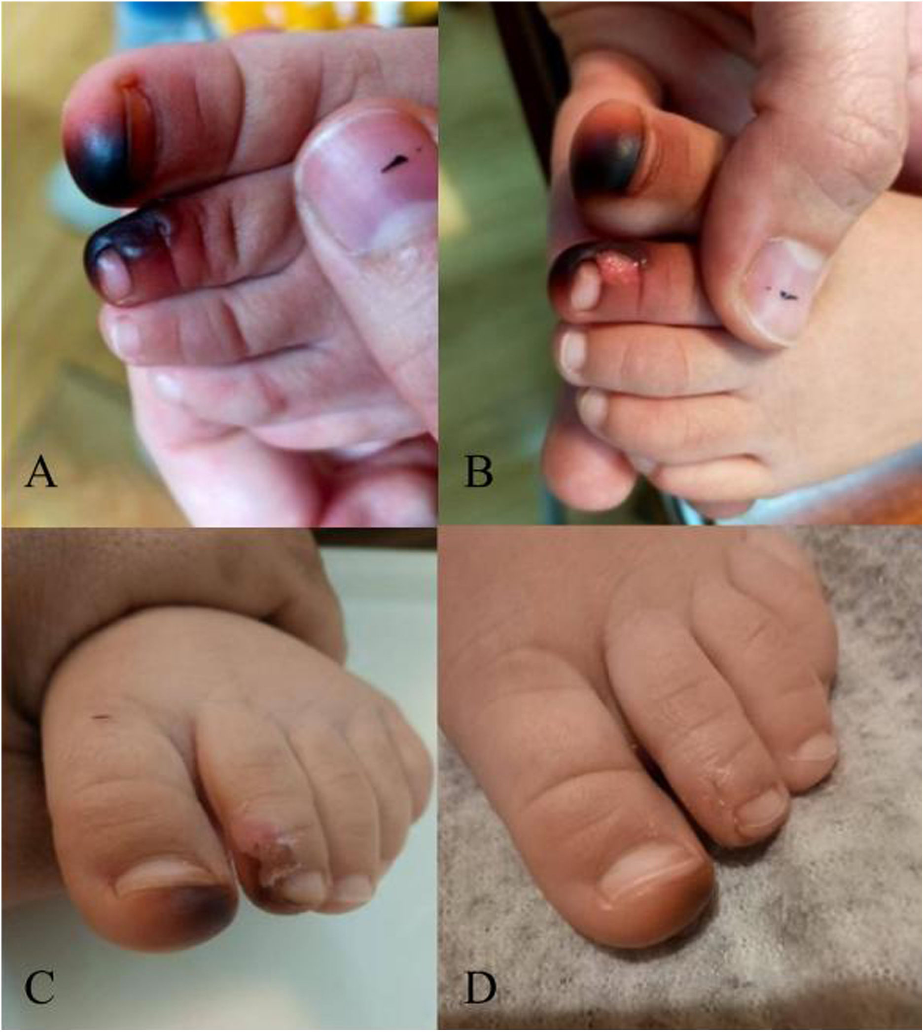
The observed lesions were consistent with those caused by the Blaps lusitanica beetle, of the Coleoptera order and Tenebrionidae family. On account of the limited experience with this type of lesion and the presence of a blister, the patient was discharged home with a prescription for a 1-week course of oral amoxicillin-clavulanic acid, and the pigmentation resolved within 2 weeks.

Similar cases have been described in the Mediterranean region.1–3 When threatened, the beetle secretes a dark and malodorous substance rich in carbohydrates and quinines. It usually only stains the skin, and in some instances also causes blisters or dermatitis. The differential diagnosis of acral purpuric skin lesions in children includes traumatic haemorrhage under the stratum corneum, vasospasm, as seen in Raynaud disease or other connective tissue diseases, Janeway lesions characteristic of bacterial endocarditis, cryoglobulinaemia, thromboangiitis obliterans, or acral melanoma. A detailed history-taking and exhaustive physical examination are essential to identify the insect as the cause and avoid performance of additional diagnostic tests.

Conflicts of interest

The authors have no conflicts of interest to declare.

References
[1]
M. Álvarez-Salafranca, O. Yélamos, M. Ramírez-Lluch, A. Valero-Torres, M. Ara-Martín.
Exogenous acral pigmentation induced by coleoptera: an underdiagnosed mimic of severe disease.
Dermatol Online J [Internet], (2020),
[2]
S. Mokni, L. Boussofara, W. Saidi, A. Aounallah, C. Belajouza, N. Ghariani, et al.
Four cases of exogenous acral pigmentation related to a darkling beetle (coleoptera: tenebrionidae: blaps).
J Eur Acad Dermatol Venereol [Internet]., 31 (2017), pp. e330-331
[3]
J. Arredondo Montero, M. Román Moleón, M.D.C. Sarmiento, G. Antona, M. Miguel-Ferrero, N. Martín-Calvo.
Exogenous acral pigmentation secondary to beetle exposure.
Pediatr Dermatol [Internet], 38 (2021), pp. 1594-1596
Copyright © 2023. Asociación Española de Pediatría
Idiomas
Anales de Pediatría (English Edition)
Article options
Tools
es en

¿Es usted profesional sanitario apto para prescribir o dispensar medicamentos?

Are you a health professional able to prescribe or dispense drugs?