Journal Information
Vol. 98. Issue 5.
Pages 389-390 (01 May 2023)
Vol. 98. Issue 5.
Pages 389-390 (01 May 2023)
Images in Paediatrics
Full text access
Newborn with elbow lump
Recién nacido con tumoración en el codo
Visits
1164
Álvaro Cobreros-Péreza,
Corresponding author
alvaro_cobre@msn.com

Corresponding author.
, Eva Pueyo-Agudob, Laura Almendro-Garcíac, María Jurado-Tabaresc
a Unidad de Gestión Clínica de Neonatología, Hospital Materno-Infantil Regional Universitario de Málaga, Málaga, Spain
b Unidad de Gestión Clínica de Pediatría, Hospital Materno-Infantil Regional Universitario de Málaga, Málaga, Spain
c Unidad de Cirugía Pediátrica, Hospital Materno-Infantil Regional Universitario de Málaga, Málaga, Spain
This item has received
Article information
Full Text
Bibliography
Download PDF
Statistics
Figures (3)
Show moreShow less
Full Text

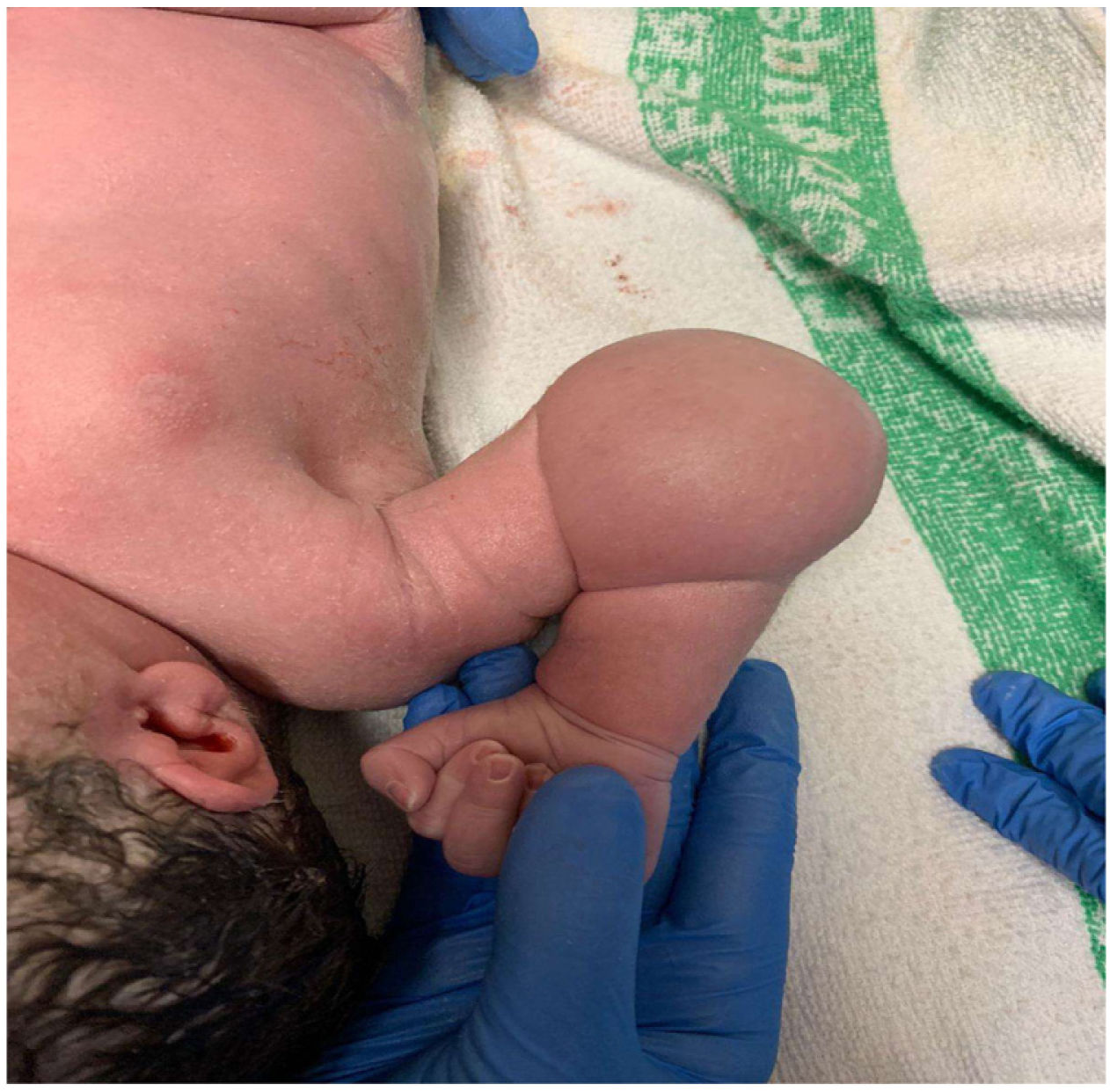
Male neonate born at 37 weeks of gestation, with no obstetric or maternal history of interest. The first physical examination revealed a mass in the right elbow (Fig. 1), with no other abnormalities.

Figure 1.

Mass in right elbow: soft mass measuring approximately 5 × 5 cm, unattached to deeper structures, absence of erythema or other cutaneous signs of inflammation.

(0.21MB).

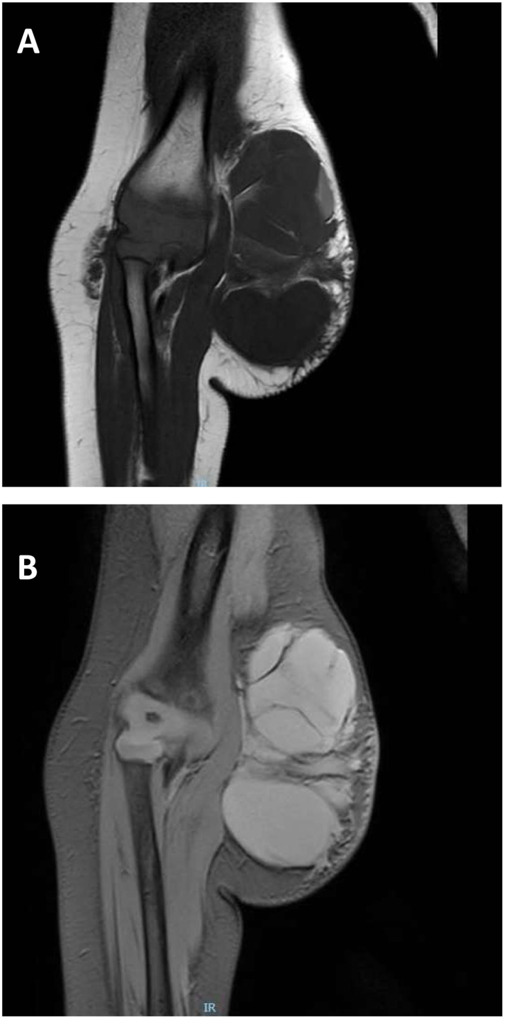
The infant was assessed in the paediatric surgery and radiology departments (Figs. 2 and 3), leading to diagnosis of a microcystic lymphatic malformation.1–3 Following a multidisciplinary evaluation, the decision was made to surgically remove the lesion with intraoperative neurophysiological monitoring at age 1.5 years. The histological examination of the specimen confirmed the suspected diagnosis, with detection of the D2 40 marker (+). No further abnormalities have been identified in the follow-up to date.

Figure 2.

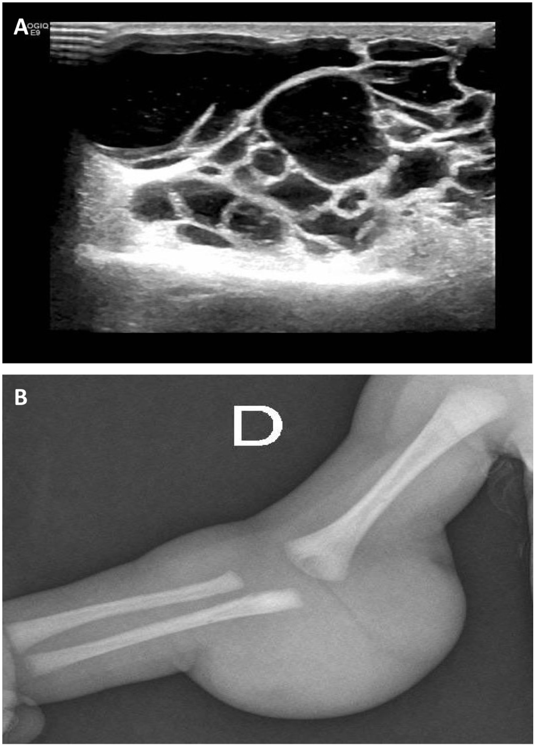
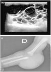
Ultrasound scan and plain radiograph of the lesion.

(A) Ultrasound scan of the right upper extremity: multicystic lesion measuring 52 × 34 × 52 mm (CC × AP × TR), predominantly avascular, located in the subcutaneous tissue of the posterior elbow and extending into the adjacent compartments. No involvement of bone, joints or muscle tissue. (B) Plain radiograph of the right upper extremity showing a tumour in the right elbow.

(0.25MB).
Figure 3.

Magnetic resonance imaging of the lesion.

(A) Fat-suppressed proton density-weighted T1-weighted image. (B) Gradient echo T2-weighted image.

(0.27MB).

Lymphatic malformations, formerly known as lymphangiomas, are congenital malformations resulting from the obstruction of lymphatic vessels, usually in the cervical region.1,3 It is rarely found in the upper extremities. Sonography2 is useful for their diagnosis, as it can detect the presence of cysts. They rarely progress to malignant tumours. Watchful waiting is a controversial approach, as these masses rarely regress spontaneously.1 There are several options for medical management2,3 (sirolimus, OK-432 or doxycycline) reserved for macrocystic malformations. However, surgical management1–3 continues to be the most frequent approach, although the appropriate timing is still under debate: some authors recommend surgery at birth, and others after 1–2 years of life.

References
[1]
M.J.H. Rahal, M.R. Abou Al Ezz, R.A. El Hajj, J.Z. Chokor, S.M. Nasser, R.C. Moucharafieh.
Elbow cystic lymphangioma in an 8-month-old boy.
Case Rep Orthop., 2019 (2019),
[2]
H. Hassan, K. AbdElelah Aly.
Management of cystic lymphangioma.
Ann Pediatr Surg, 8 (2012), pp. 123-128
[3]
C. Damaskos, N. Garmpis, M. Manousi, A. Garmpi, G.A. Margonis, E. Spartalis, et al.
Cystic hygroma of the neck: single center experience and literature review.
Eur Rev Med Pharmacol Sci., 21 (2017), pp. 4918-4923
Copyright © 2022. Asociación Española de Pediatría
Idiomas
Anales de Pediatría (English Edition)
Article options
Tools
es en

¿Es usted profesional sanitario apto para prescribir o dispensar medicamentos?

Are you a health professional able to prescribe or dispense drugs?