


Testicular tumours are infrequent in the paediatric age group. The most frequent type are germ cell tumours.1 Juvenile granulosa cell tumour (JGCT) are rare sex-cord stromal tumours with an estimated incidence of 0.5 cases per 100 000 children.2
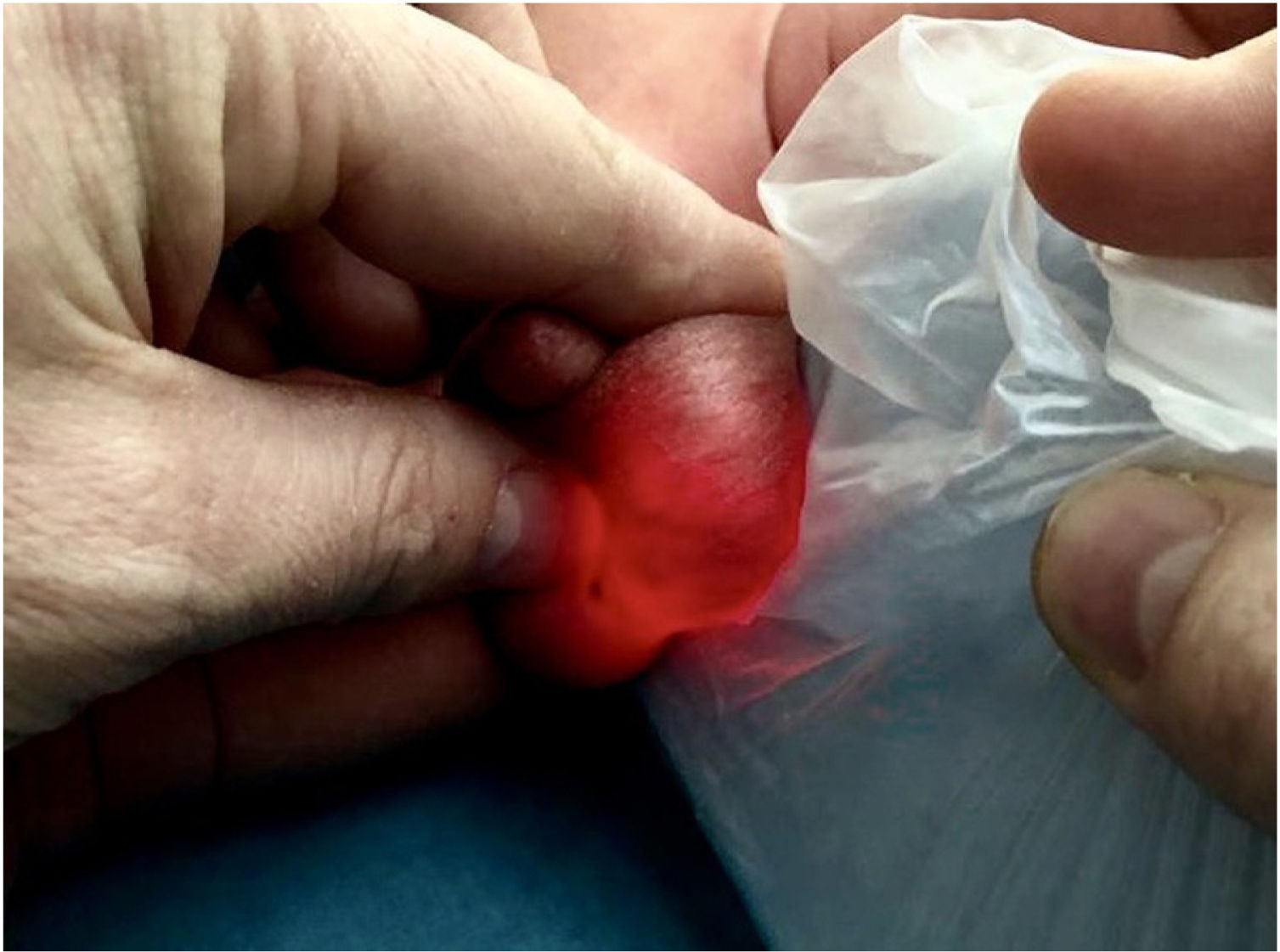
We present the case of a male newborn in whom prenatal ultrasound examinations were normal that had testicular asymmetry at birth with enlargement of the left testis. Transillumination allowed discernment of septations within the scrotal sac (Fig. 1).

The ultrasound examination found a multicystic mass in the left testicle (Fig. 2), so the evaluation was completed with assessment of tumour markers: alpha-foetoprotein (AFP) >20 000 ng/mL; beta human chorionic gonadotropin (HCG) <1.2 mU/mL (negative); testosterone 0.3 ng/mL and inhibin B 72 pg/mL, findings suggestive of JGCT.

The differential diagnosis includes other diseases that manifest with cystic lesions in the testis, such as testicular cystic teratoma or yolk sac tumour.3
The patient underwent a radical orchiectomy at 35 days post birth with a favourable outcome and normalization of AFP levels by age 9 months.
The gross and histological examinations found a lesion confined to the testis with a solid follicular pattern lined with granulosa cells with immunostaining positive for expression of vimentin, inhibin, CD99, CKAE1/AE3 and S-100 (Fig. 3).

Juvenile granulosa cell tumour manifests as a painless scrotal mass and involves the left testis more frequently. There are no reported cases of recurrence or metastasis.3 Orchiectomy is considered the curative treatment. In some cases, JGCT is associated with malformation of the urogenital system or sex chromosome anomalies.2
FundingThis research did not receive any external funding.
Conflicts of interestThe authors have no conflicts of interest to declare.
We thank Margarita Llavador Ros, of the Department of Pathological Anatomy, Hospital Universitario y Politécnico La Fe, Valencia, Spain.

