


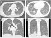
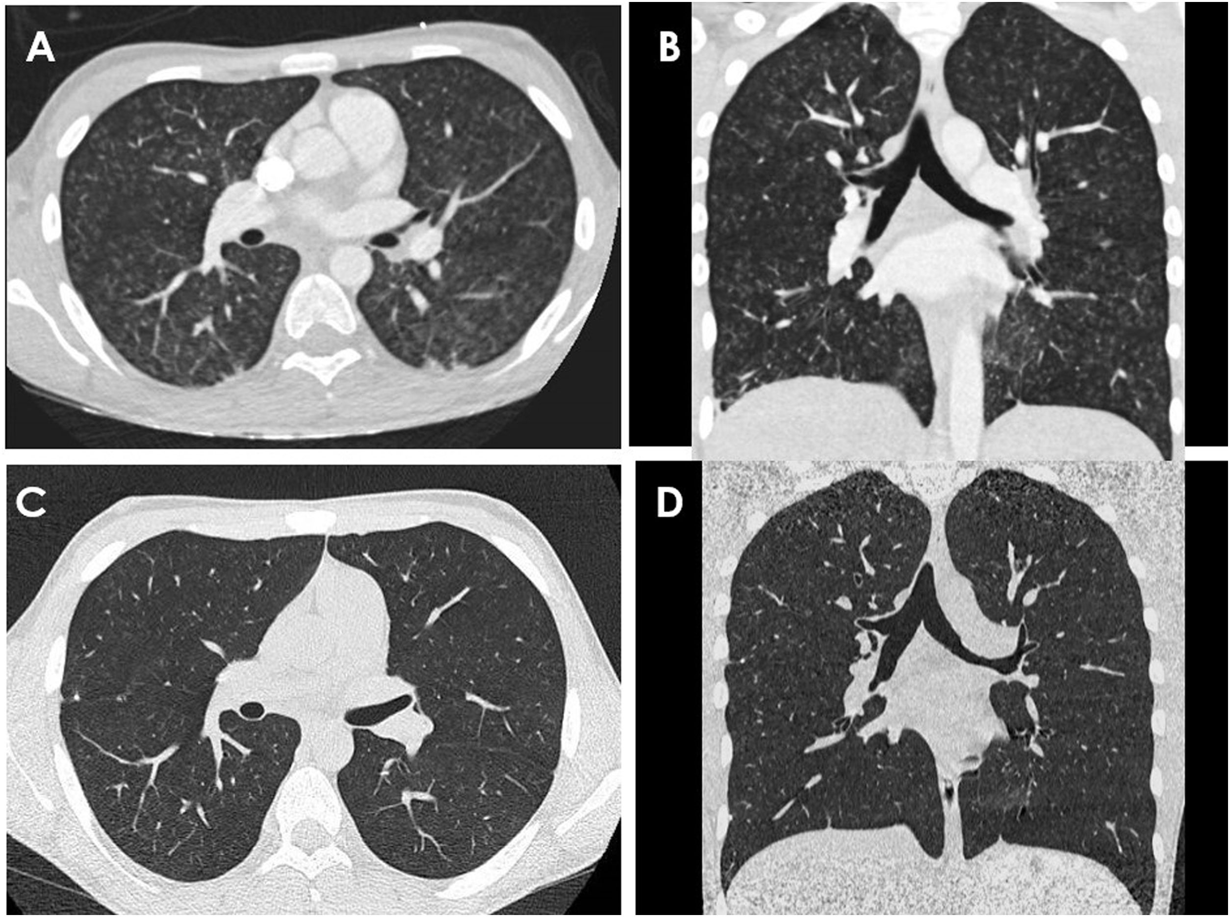
We describe the case of a male adolescent aged 17 years admitted for severe acute respiratory failure with hypoxaemia. During the hospital stay, numerous diagnostic tests were performed, and the findings of the computed tomography scan of the lungs proved key to the diagnosis, with visualization ground glass micronodules with tree-in-bud opacities and a diffuse distribution1 (Fig. 1).
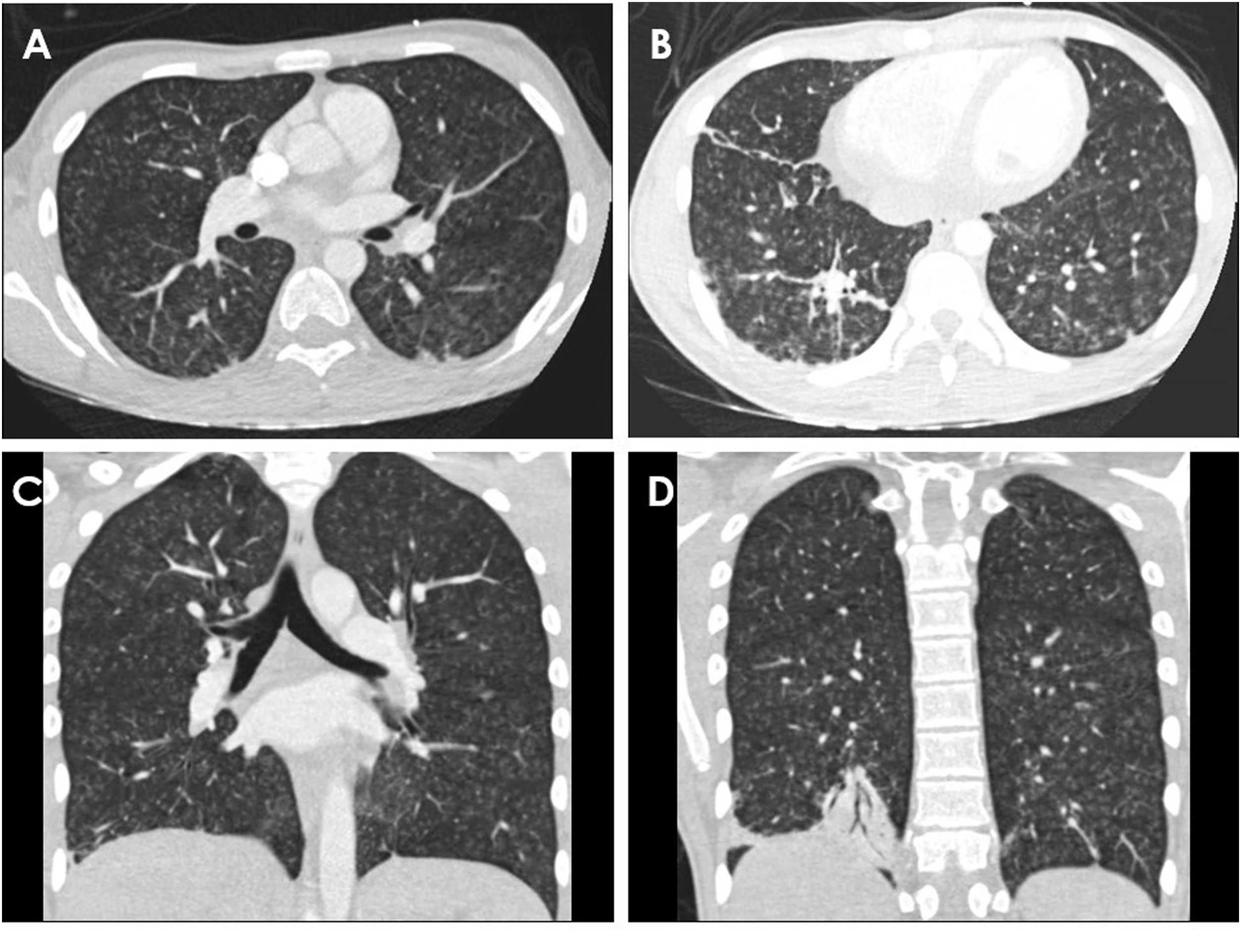
Computed tomography images obtained during the hospital stay. Axial (A and B) and coronal (C and D) images showed small micronodular opacities in a diffuse tree-in-bud pattern, without involvement of the subpleural region. Images B and D evince the presence of consolidations in the right lower lobe.
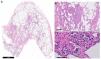
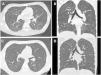
With these features, the most likely diagnosis is acute pneumonitis of infectious, autoimmune or inflammatory aetiology; the first 2 possibilities were ruled out thoroughly and to explore the possibility of inflammation, we chose to perform a video-assisted thoracoscopic lung biopsy (Fig. 2), and the resulting findings were compatible with subacute hypersensitivity pneumonitis. The patient had not been exposed to any of the classical triggers (birds, moulds, chemicals …) but he had engaged in an increasing consumption of cannabis resin, which he smoked in the weeks preceding the onset of symptoms.2 The patient was treated with steroids at tapering doses over 5 months and instructed not to consume smoked or inhaled substances, with clinical and radiological evidence of improvement 6 months later3 (Fig. 3).
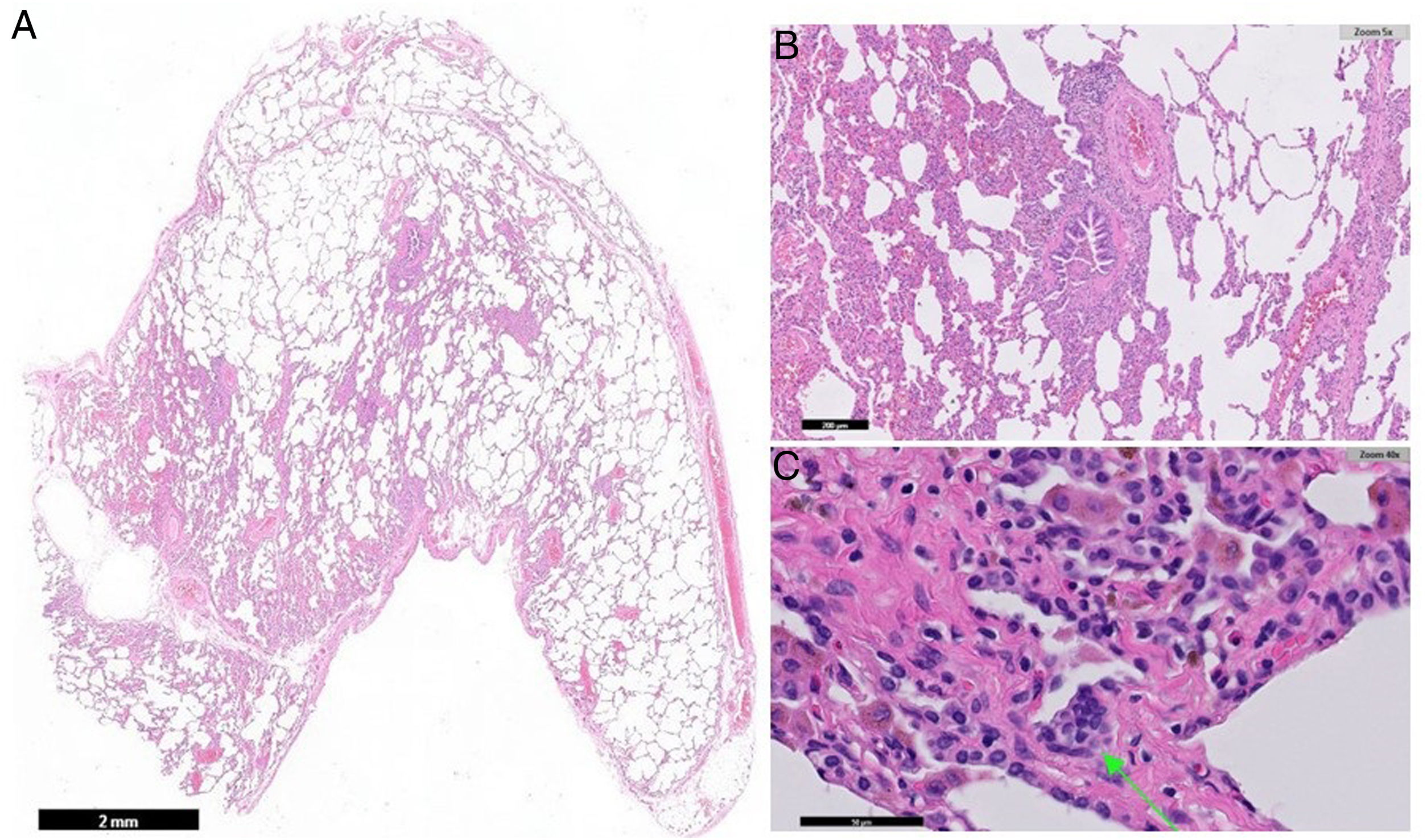
Histological sections of the biopsy specimen of the right lower lobe of the lung (images A, B and C). Image A (0.5×) presents a panoramic view of the lung wedge resection specimen with partial architectural distortion and presence of nodular structures. Lymphoplasmacytic and histiocytic infiltration in the lung parenchyma in the bronchovascular axis and accumulation of alveolar macrophages B y C). The arrow points at a cluster of histiocytes compatible with a multinucleated giant cell (C).
We considered reporting this case relevant due to the alarming increase in the consumption of recreational drugs by the adolescent population at increasingly early ages.
Previous meeting: This case was presented at the XLIII Meeting of the Sociedad Española de Neumología Pediátrica (SENP) entering the competition for the clinical case awards, receiving the second prize.
