


Up to 60% of hospitalised neonates may develop incontinence-associated dermatitis (IAD). Our aim was to adapt the Clinical Evaluation Scale for Characterization of the Severity of Diaper Dermatitis to the Spanish population and to find out the nationwide frequency of IAD in hospitalized neonates.
MethodsCross-cultural adaptation and assessment of content validity of the scale. We carried out a prospective, multicentre observational study of the incidence of nappy rash in postnatal wards and neonatal intensive care units in 6 Spanish hospitals.
ResultsWe obtained a content validity index of 0.869 for the total scale (95% CI, 0.742−0.939). The sample included 196 neonates. The cumulative incidence of IAD was 32.1% (9.1% mild-moderate, 8% moderate and 1.6% severe). The incidence rate was 2.2 IAD cases per 100 patient days. A stool pH of less than 5.5, a greater number of bowel movements a day, a greater daily urine output and the use of oral drugs were among the factors associated with the development of IAD.
ConclusionThe Spanish version of the Clinical Evaluation Scale for Characterization of the Severity of Diaper Dermatitis had an adequate content validity for the assessment of DAI in the hospitalised neonatal population. Mixed feeding, treatment with oral drugs and the use of medical devices in the perianal area were associated with an increased risk of nappy dermatitis in infants.
Hasta el 60% de los neonatos hospitalizados pueden desarrollar una dermatitis asociada a la incontinencia (DAI). El objetivo fue adaptar al contexto español la escala Clinical evaluation scale for characterization of the severity of Diaper Dermatitis y averiguar la magnitud nacional de la DAI en neonatos hospitalizados.
MetodologíaAdaptación transcultural y validez de contenido. Estudio observacional, prospectivo y multicéntrico de incidencia de dermatitis del pañal en unidades de hospitalización y cuidados intensivos neonatales de 6 hospitales españoles.
ResultadosSe obtuvo un índice de validez de contenido de 0.869 para el total de la escala (IC95% 0.742 – 0.939). Se incluyen un total de 196 neonatos. Se obtuvo una incidencia acumulada de DAI del 32.1% (9.1% leves-moderadas, 8% moderadas y 1.6% grave). La tasa de incidencia fue de 2.2 DAI por cada 100 neonatos/día de ingreso. El pH de las heces <5.5, el nº deposiciones/día, los ml de orina/día y los fármacos orales fueron algunos de los factores favorecedores de DAI.
ConclusionesLa versión española de la Clinical evaluation scale for characterization of the severity of Diaper Dermatitis tiene un contenido válido para valorar DAI en población neonatal ingresada. Los neonatos portadores de lactancia mixta, fármacos orales y dispositivos locales en la zona perianal tienen más riesgo de presentar dermatitis del pañal.

In newborns, incontinence-associated dermatitis (IAD), commonly known as diaper dermatitis (DD) or nappy rash, is an inflammatory irritant lesion that manifests in areas covered by the diaper or adjacent to it.1 This irritant dermatitis is a serious problem resulting from the particular combination of moisture, maceration, friction and contact with urine, stools and other substances during the neonatal period.1,2
The development of DD is attributed to the impairment of the barrier properties of the skin in addition to the irritants themselves.3 The action of external irritants (faeces, urine, diaper material, moisture, friction)4,5 and internal factors (changes in skin pH, excessive transepidermal water losses, excessive hydration of the skin and low zinc levels)3,6 promote the development of lesions, which tend to be complicated by the action of the microorganisms present in the area.5,7,8
The incidence of IAD in the neonatal population in the community is estimated at 25%–70%.9–12 In hospitalized newborns, the reported incidence also varies widely between studies, ranging from 4.7% to 60%.13–17
The variation among studies is not limited to the reported outcomes, but also extends to the methods used to assess IAD (clinical judgment of dermatologists, clinical judgment of health care providers or use of standardised assessment scales).13–17 Stamatas et al. recommended the use of IAD severity rating scales specific to the paediatric population, as opposed to the scales used widely in the scientific literature that were developed for use in the adult population.18,19
Diaper dermatitis is a frequent iatrogenic adverse event in infants that warrants investigation of its magnitude as well as of the best methods for its clinical assessment and prevention.10,19 The lack of multicentre epidemiological studies and tools for assessment of IAD validated for use in the paediatric and neonatal population in our health system motivated us to adapt the instrument recommended by Stamatas et al. and determine the nationwide incidence of IAD in hospitalised newborns in Spain, in addition to assessing the risk factors and preventive measures associated with its development.
MethodsTranslation and cultural adaptation of the Clinical Evaluation Scale for Characterization of the Severity of Diaper DermatitisAfter obtaining the permission of the authors of the original scale,18 we carried out its cross-cultural adaptation through the forward and back translation process, carried out by two independent translation companies. The research team developed a version of the scale in a consensus process without adding any concept that was not already present in the translations. Subsequently, this consensus-based version was back translated by two bilingual translators to assess the equivalence of its items to those of the original scale.
Content validity of the Clinical Evaluation Scale for Characterization of the Severity of Diaper DermatitisWe assessed content validity by calculating the content validity index (CVI) for each item applying 4 criteria: ambiguity, simplicity, clarity and relevance. Each of these criteria were rated on a Likert scale going from 1 to 4. These assessments were made by a panel of 15 experts20 appointed through an adaptation of the method proposed by Fehring21 (health care provider specialised in paediatric care [3 points], having a master’s or doctorate degree [1 point], having written a dissertation relevant to the subject under study [2 points], having published one or more articles relevant for the subject matter in peer-reviewed journals [2 points], belonging to a recognised research group or experience in research relevant to the subject matter [2 points], having a minimum of 5 years’ experience in paediatric care practice [2 points] or a minimum of 1 year’s experience in neonatal care [2 points]). A minimum of 5 points were required to be considered an expert. We consulted experts by extending an invitation to participate to all experts via electronic mail, including a link to the online questionnaire (LimeSurvey software package: http://limesurvey.org).
Incidence of diaper rashWe conducted a multicentre, prospective and observational incidence study of DD in 6 neonatal units in Spain (Hospital Clínico de Valencia, Hospital Clínico San Carlos de Madrid, Hospital General Universitario de Valencia, Hospital Fundación Jiménez Díaz de Madrid, Hospital Son Llàtzer de Palma de Mallorca y Hospital San Pedro de Logroño) between March and July of 2016. We excluded neonates with a length of stay of less than 72 h, neonates who already had IAD or lesions in the perianal region and neonates who had received a diagnosis of dermatological disease. To assess IAD, we used the Spanish version of the Clinical Evaluation Scale for Characterization of the Severity of Diaper Dermatitis, defining cases of dermatitis as patients who developed lesions of at least “mild” degree.
Sample size and statistical analysisThe CVI was calculated using the Aiken V coefficient, defining content validity as a value of 0.75 or greater.20 To analyse the confidence intervals of the Aiken V coefficient, we used a free software tool developed with the Visual Basic 6.0 language (Microsoft, WA, USA).22
To calculate the sample size for the incidence study, we used an expected proportion of DD of 0.3 for a 95% confidence level, a precision of 0.08 and estimated losses of 10%, which yielded a sample size of 140 participants to be distributed among the 6 hospitals in proportion to their volume of admissions.
We performed a descriptive analysis of the sample and estimated the incidence rate and cumulative incidence of IAD. We used the χ2 test or Fisher exact test and the Student t test or Mann-Whitney U test as applicable, and carried out a one-way analysis of variance (ANOVA) with the post hoc Bonferroni (homogeneous variance) or Games-Howell correction (heterogeneous variance) to assess for differences between the explanatory variables potentially associated with the development of IAD. For variables that were significantly associated with IAD, we calculated the odds ratio (OR) to determine the degree to which they behaved as risk factors/preventive factors in relation to IAD. Lastly, we fitted a logistic regression model with the variables that had exhibited significant differences. The data were tabulated and analysed with the statistical package SPSS version 21.0 (IBM Corp Armonk, NY, USA; 2012). We set the level of significance at 0.05.
Control for biasUniformity in data collection was promoted by holding training sessions on how to use the Spanish version of the Clinical Evaluation Scale for Characterization of the Severity of Diaper Dermatitis. In addition, all health care professionals in the participating neonatal units received training on data collection. The audiovisual resources used for training were hosted in the website www.upppediatria.org.
ResultsCross-cultural adaptation and content validity of the Clinical Evaluation Scale for Characterization of the Severity of Diaper DermatitisAfter the forward and back translation process, we obtained the definitive version in Spanish of the scale (Appendix A supplemental material).
A panel of 15 experts carried out the content validation of the Spanish version of the scale. The analysis of the data yielded an index of 0.869 for the total scale (95% CI, 0.742−0.939), and all items were considered valid (Table 1).
Content validity of the Spanish version of the Clinical Evaluation Scale for Characterization of the Severity of Diaper Dermatitis.
| Item | Aiken’s V | 95% CI |
|---|---|---|
| Normal skin | 0.963 | 0.862−0.990 |
| Very mild | 0.791 | 0.652−0.884 |
| Mild | 0.861 | 0.731−0.993 |
| Mild/moderate | 0.868 | 0.739−0.938 |
| Moderate | 0.833 | 0.699−0.914 |
| Moderate/severe | 0.840 | 0.707−0.919 |
| Severe | 0.930 | 0.817−0.975 |
| Total scale | 0.869 | 0.742−0.939 |
CI, confidence interval.
The study included 196 neonates. Of this total, 53.6% were male, 81.1% were white, 57.7% were born preterm and 51.5% had a history of complicated delivery.
Perinatal conditions accounted for 55.1% of the principal diagnoses in the sample, followed by endocrine or nutritional disorders (12.8%).
Incidence of diaper dermatitisWe found a cumulative incidence of IAD of 34.7%. The incidence rate was 2.9 cases of IAD per 100 neonatal inpatient days. The mean inpatient days elapsed to the first episode of dermatitis was 10.3 (SD, 10).
Based on the assessment with the Clinical Evaluation Scale for Characterization of the Severity of Diaper Dermatitis, 13.2% of the cases were mild to moderate, 11.8% were moderate and 1.5% were severe. In 70.6% of cases, IAD involved the perianal area, in 8.8% the buttocks and in 7.4% the groin. We assessed for the presence of local infection in newborns with IAD; pathogens were not isolated in 82.4% of episodes and bacterial pathogens were detected in the remaining 17.6% restante. Fungal pathogens were not isolated in any of the patients.
Table 2 summarises the characteristics of the patients with and without IAD.
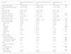
Description of the sample based on the presence or absence of IAD.
| Variables | IAD (n = 68) | No IAD (n = 128) | P |
|---|---|---|---|
| Sex | 47.1% male; 52.9% female | 57% male; 43% female | .183 |
| Gestational age | 35.5 [± 3.9] | 35.3 [± 4] | .769 |
| Birth weight | 2.426.0 [± 870.4] | 2.336.6 [± 867.7] | .493 |
| Birth length | 44.4 [± 7.4] (n = 67) | 44.3 [± 5] | .921 |
| Length of stay (days) | 16.9 [± 14.5] | 12.5 [± 13.4] | .037 |
| Age at discharge (days post birth) | 20 [± 15.4] | 15.6 [± 13.8] | .039 |
| Bowel movements/day | 4.0 [± 1.7] (n = 62) | 2.9 [± 1.2] (n = 125) | .000 |
| Mean faecal PH during stay | 5.9 [± 0.8] (n = 62) | 6 [± 0.5] (n = 125) | .240 |
| Mean faecal PH < 5.5 or > 7 | 39.7% (n = 62) | 25% (n = 125) | .033 |
| Mean daily urine volume (mL) | 249.03 [± 105.1] (n = 63) | 219.6 [± 92.4] (n = 109) | .069 |
| Mean urine pH | 6.2 [± 0.6] (n = 66) | 6.1 [± 0.4] (n = 127) | .153 |
| Mean urine pH < 5.5 or > 7 | 16.2% (n = 66) | 18% (n = 127) | .752 |
| Detection of nitrites and/or bilirubin in urine | 28% | 27.5% | .841 |
| Urinary catheter | 5.9% | 3.9% | .382 |
| Orogastric tube | 58.8% | 51.6% | .332 |
| Nil per os | 33.8% | 28.1% | .408 |
| Feeding modality | |||
| Exclusive formula feeding | 29.4% | 34.4% | .294 |
| Exclusive breastfeeding | 10.3% | 31.3% | .001 |
| Mixed feeding | 60.3% | 34.4% | .000 |
| Parenteral nutrition | 26.5% | 29.7% | .635 |
| Collection bag | 29.4% | 11.7% | .002 |
| Oral pharmacotherapy | 64.7% | 43% | .004 |
| Vitamin D3 | 69.1% | 45.3% | .001 |
| Iron | 11.8% | 10.2% | .729 |
| Zinc sulphate | 0% | 0% | – |
| Antifungal | 5.9% | 3.9% | .529 |
| Antibiotic | 1.5% | 0.8% | .648 |
| Sucrose | 17.6% | 8.6% | .061 |
| Multivitamin | 7.4% | 0.8% | .011 |
| Other | 0% | 4.4% | .041 |
| IV antibiotherapy | 39.7% | 37.5% | .762 |
| Skin barrier products | 82.4% | 3.1% | .000 |
| Cleansing method | |||
| Soft pad + tap water | 54.0% | 44.4% | .208 |
| Cotton gauze + tap water | 15.9% | 13.5% | .662 |
| Cotton gauze + sterile water | 7.9% | 5.3% | .466 |
| Soft pad + cleansing product | 0% | 0.8% | .679 |
| Commercial wipe | 22.2% | 36.1% | .051 |
| Kangaroo care | 72.1% | 72.4% | .955 |
| Frequency of diaper change | |||
| Every/2 h | 16.4% | 20.8% | .821 |
| Every 3 h | 76.1% | 72.9% | |
| Every 4 h | 7.5% | 6.3% | |
| Type of resting surface | |||
| Static SSMP | 91% | 81.1% | .069 |
| Dynamic SSMP | 9% | 18.9% |
SSMP, special surface to manage pressure.
Bold entries are just to remark statistical significances differences.
As regards the protective effect of breastfeeding and the association of mixed feeding with the risk of IAD, we made a descriptive analysis of the characteristics of the patients based on the feeding modality during the hospital stay for their comparison (Table 3).
Description of the patients in the sample by feeding modality.
| Variables | Exclusive breastfeeding (n = 64) | Exclusive formula feeding (n = 47) | Mixed feeding (n = 85) | P |
|---|---|---|---|---|
| Length of stay (days) | 11.52 ± 13.85 | 15.04 ± 11.52 | 15.41 ± 15.07 | .206 |
| Time of discharge (days post birth) | 14.75 ± 14.33 | 18.60 ± 10.88 | 18.11 ± 16.31 | .278 |
| Gestational age | 36.47 ± 3.65a | 33.55 ± 4.58a,b | 35.67 ± 3.47b | |
| Mean faecal pH | 5.87 ± 0.77a | 6.30 ± 0.52a,c | 5.97 ± 0.85c | |
| Faecal pH < 5.5 or > 7 | 34.3% | 19.1% | 32.9% | .169 |
| Bowel movements/day | 3.10 ± 1.49 | 2.97 ± 1.42 | 3.59 ± 1.64 | .054 |
| Mean urine pH media | 6.97 ± 0.74d,e | 6.14 ± 0.69d | 6.63 ± 0.86e | |
| Mean urine pH < 5.5 or > 7 | 20.3% | 14.9% | 16.5% | .728 |
| Urine output (mL/day) | 241.34 ± 103.57 | 198.35 ± 116.14 | 236.42 ± 80.65 | .090 |
| Oral pharmacotherapy | 40.6% | 57.4% | 54.1% | .146 |
| Vitamin D3 | 46.9% | 55.3% | 57.6% | .411 |
| Iron | 6.3% | 17% | 10.6% | .190 |
| Antifungal | 3.1% | 0% | 0% | .0124 |
| Antibiotic | 0% | 10.6% | 4.7% | .614 |
| sucrose | 10.9% | 8.5% | 14.1% | .176 |
| Multivitamin | 4.7% | 0% | 3.5% | .301 |
| Other | 0% | 2.1% | 2.4% | |
| Collection bag | 12.5% | 8.5% | 27.1% | .011 |
| Cleansing method | ||||
| Soft pad + tap water | 31.3% | 76.6% | 43.5% | .000 |
| Cotton gauze + tap water | 6.3% | 2.1% | 27.1% | .000 |
| Cotton gauze + sterile water | 9.4% | 2.1% | 5.9% | .288 |
| Soft pad + cleansing product | 0% | 2.1% | 0% | .203 |
| Commercial wipe | 53.1% | 17% | 23.5% | .000 |
| Type of SSMP | ||||
| Static SSMP | 95.3% | 59.6% | 90.4% | .000 |
| Dynamic SSMP | 4.7% | 40.4% | 9.6% |
Exclusive breastfeeding: infant exclusively breastfed throughout the hospital stay; exclusive formula feeding: infant fed formula exclusively during the stay; mixed feeding: infant fed human milk and formula at different points of the stay.
a,b,c,d,eStatistical significance in ANOVA with post hoc Bonferroni/Games–Howell correction.
Bold entries are just to remark statistical significances differences.
The odds ratios for the presence of IAD for the variables found to be significantly associated with its development are presented in Table 4.
Odds ratio for the presence of IAD in relation to significantly associated variables.
| Variables | OR | 95% CI | P |
|---|---|---|---|
| Length of stay (days) | 1.02 | 1.001−1.043 | .041 |
| Age at discharge (days post birth) | 1.02 | 1.001−1.041 | .043 |
| Faecal pH < 5.5 or > 7 | 1.97 | 1.053−3.707 | .034 |
| Bowel movements/day | 1.59 | 1.281−1.997 | .000 |
| Exclusive breastfeeding | 0.25 | 0.106−0.601 | .002 |
| Mixed feeding | 2.89 | 1.579−5.322 | .001 |
| Collection bag | 3.13 | 1.483−6.644 | .003 |
| Oral pharmacotherapy | 2.43 | 1.325−4.470 | .004 |
| Skin barrier products | 144.66 | 44.686−468.339 | .000 |
CI, confidence interval; OR, odds ratio.
We fitted a model for the risk of neonatal IAD that explained 50% of the observed variance (Table 5).
DiscussionWe undertook a study for the Spanish translation and cross-cultural adaptation of the Clinical Evaluation Scale for Characterization of the Severity of Diaper Dermatitis,18 with a subsequent assessment of its content validity, leading to the conclusion that the Spanish version is a valid instrument to grade DD in the neonatal population of Spain. A literature review conducted by Dunk et al.19 in 2022 identified as many as 8 instruments used for the assessment of DD in the paediatric population in different studies, which results in inconsistencies in the language and criteria applied to its definition and grading and hinders the comparison of data from different studies.23 In Spain, the Clinical Evaluation Scale for Characterization of the Severity of Diaper Dermatitis is the first instrument validated for the neonatal population. In their reviews, Esser and Dunk concluded that the availability of validated scales would allow the standardization of definition criteria, measurement of the frequency of IAD, assessment of its severity and evaluation of treatment outcomes.19,23 The heterogeneity of the instruments used for the same purpose, validated in different countries and applicable to different age groups makes it difficult to generalise research findings. International initiatives like the Ghent Global Incontinence Associated Dermatitis Categorization Tool (GLOBIAD), validated in 30 countries for use in the adult population, exemplifying how validation processes should be undertaken in the future. The use of a single scale, modified, adapted and validated in both the adult and paediatric populations would facilitate its clinical application through the standardization of diagnostic criteria and the effective management of the preventive resources available in health care facilities.24,25
Applying the dermatitis severity rating scale in multiple centres, we found that 3 out of 10 hospitalised neonates had some degree of IAD. The incidence of IAD reported by studies in hospitalised neonates is variable. The studies of Meszes et al.16 and Csoma et al.,15 in which dermatitis was assessed by a dermatologist, found an incidence of 4.7% (n = 211) and 5.4% (n = 460), respectively. Visscher et al.,14 in a study in which the assessment was made by nurses using standardised numerical scales for erythema and dryness and physiological measures, found an incidence of 60% (n = 130). Alonso et al.13 reported an innocence of 19.7% (n = 213) based on the assessment by paediatricians and the application of the scale proposed by Lane and Drost modified by Kiechl-Kohlendorfer. Malik et al.17 carried out a retrospective in 1241 hospitalised newborns and found an incidence of DD of 23% based on the documentation in the health records. The heterogeneity in the assessment approaches hinders the comparison of our findings with those of previous studies. The high percentage of cases categorised as mild (71.4%) may not have been considered IAD in assessments based on the clinical judgment of the provider. We consider that early detection of this type of lesions is important in clinical practice, for, as our findings illustrate, preventive measures like the use of barrier products (containing zinc oxide, silicone or polyurethane, fatty acids, etc) behaved as risk factors in our analysis because they were not implemented until the rash was detected.
As regards the factors associated with IAD in our study, gestational age, sex or birth weight were not significantly associated with it. This was consistent with the findings of previous studies in which the fragility of the skin of preterm infants did not seem to be associated with an increase in IAD, and the frequency of IAD episodes was higher with increasing age in weeks.13,14,26 In our sample, the length of admission and the chronological age at discharge were significantly greater in the IAD group. In the previous literature, this direct correlation between the weeks of age and the frequency of IAD episodes has been found to be associated with an increase in the number of bowel movements and urine output,13,14,26 which in our study were also significantly greater in the IAD group.
When it came to the pH, we did not find significant differences in the mean (quantitative) faecal or urine pH during the stay between groups. Other studies considered the pH of the skin covered by the diaper14,26,27 and found a more alkaline pH, a factor that is associated with the development of IAD. Since the mean pH in urine and in faeces at admission was approximately 6, we decided to assess how many newborns had means 0.5–1 point above or below this pH value, and found that a significant percentage of newborns with dermatitis had a faecal pH of less than 5.5 or greater than 7. The damage to the skin probably depends more on significant fluctuations toward greater acidity as well as alkalinity of the eliminated waste. We did not find evidence of this association between pH and IAD for urine, which made us consider the possibility that the irritant that has an impact on the faecal pH is not at play in urine.
When we assessed the association between pharmacological treatment and the presence of dermatitis, we found an increased risk in neonates who received oral therapy (nutritional supplements, antibiotherapy, antifungal therapy, etc). While other authors had already proposed the association between antibiotherapy and the development of dermatitis,28 our findings expand the scope of the association to other types of oral drugs. We found that oral drugs of any time were used more frequently in newborns with IAD. The significantly higher frequency of IAD observed in newborns who received vitamin D supplementation may have been biased on account of age, since the age in days at discharge was significantly greater in neonates who received vitamin D supplementation compared to newborns who did not receive it (22.2 vs. 11.16 days; P = .000).
We assessed the feeding modality in neonates, and found that the frequency of dermatitis was significantly greater in those who received mixed feeding and significantly lower in those who received exclusive breastfeeding. Similarly, the study conducted by Ersoy-Evans et al.29 in a community-based sample of 63 children aged 0–24 months with dermatitis of varying severity found that those who had been breastfed had had fewer episodes of dermatitis. The authors attributed this to a lower faecal pH (5.28 in the breastfed group compared to 6.82 in the formula-fed group) and the faecal lower lipase and protease activity in the breastfed group,29 which was consistent with the findings of Berg et al.,6 who analysed frozen specimens obtained from 20 infants in the community aged 1–5 months, of who 10 were exclusively breastfed. In our study, we calculated the mean urine and faecal pH measured daily during the stay at the time of excretion, and found a significantly lower pH in the faeces of exclusively breastfed neonates. However, these infants also had a significantly higher urine pH. On the other hand, mixed feeding, a clear risk factor in the study, was not associated with significant differences in the faecal pH in comparison with the exclusive breastfeeding group. The patient age range, sample size and other conditions in the studies conducted by Ersoy-Evans et al.29 and Berg et al.6 differed significantly from our study, which could explain the observed differences. We searched for statistically significant differences based on the feeding modality groups in our sample, and found a more alkaline urine pH in the exclusively breastfed group and a higher proportion of infants who had used collection bags during the hospital stay in the mixed feeding group. In our opinion, these are relevant factors, but they cannot justify a protective effect or an increased risk associated with different feeding modalities. At present, there is no clear evidence of the association of early introduction of formula feeding or mixed feeding with other cutaneous disorders, such as atopic dermatitis.30,31 It would be interesting to explore whether the known differences or changes in in the microbiota in association with the feeding modality (differences in the concentration of short-chain fatty acids and in the composition of the microbiota)32–34 could be related to our findings.
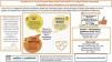
ConclusionAs newborn infants develop, the frequency of urination and bowel movements increases and this in turn increases the risk of IAD due to a greater exposure to irritants.
Newborns hospitalised in neonatal units or neonatal intensive care units with an increased stool frequency, who receive mixed-feeding, who receive oral drugs or carry medical devices in the perianal area (collection bags) are at increased risk of developing diaper dermatitis, so the preventive use of barrier products is indicated in these patients.
On the other hand, barrier products are used more frequently after the detection of dermatitis than for preventive purposes. The use of clinical prediction rules for dermatitis can help detect IAD in its early stages and promote the early use of these products to prevent progression to more severe stages.
FundingThis research project did not receive specific financial support from funding agencies in the public, private or not-for-profit sectors.
Ethical considerationsThe study was approved by the Clinical Research Ethics Committee of each participating hospital. Parents and/or legal guardians received an informational sheet and were asked to sign an informed consent form (and informed of the ability to withdraw consent at any time). The study did not include any data that could be used directly or indirectly to identify the patient, in adherence with Organic Law 3/2018, of December 5, on the Protection of Personal Data and the Guarantee of Digital Rights. The researchers in each of the participating centres were responsible for ensuring the safety of the collected data, which were to be accessed and used solely for the established purposes of the study.
We submitted the study protocol to the Agencia Española de Medicamentos y Productos Sanitarios (AEMPS, Spanish Agency of Medicines and Medical Devices), which classified it as a “non-post-authorization observational study”.
Conflicts of interestThe authors have no conflicts of interest to declare.
The following is Supplementary data to this article:
