
We present the case of a boy aged 4 years in whom a mass appeared in the right side of the neck when he cried, with onset 1 month prior to consultation. The physical examination was normal while the patient was at rest, but an elongated, soft and compressible painless mass did appear in the right side of the neck upon performance of the Valsalva manoeuvre (Appendix A, video 1).
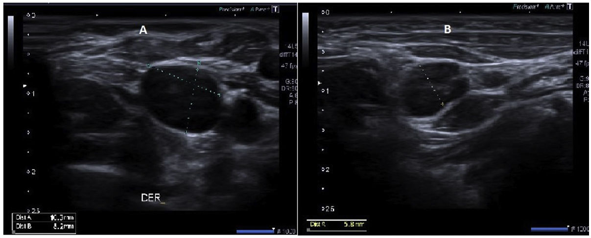
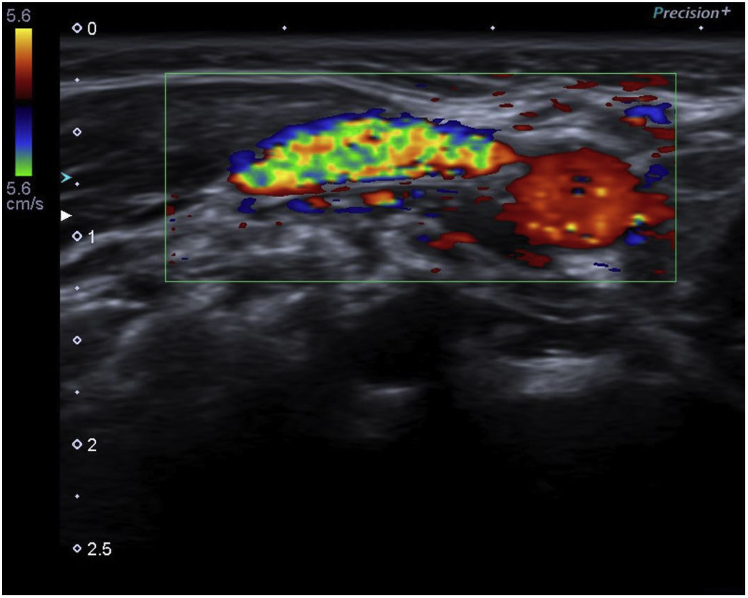
The cervical ultrasound scan showed a prominent right internal jugular vein, larger in diameter compared to the contralateral vein, that dilated with the Valsalva manoeuvre (Fig. 1). The Doppler ultrasound scan confirmed the presence of venous flow and absence of intraluminal thrombi (Fig. 2).
 Right internal jugular vein upon performance of the Valsalva manoeuvre, measuring 8.2 × 10.3 mm. (B) Left jugular vein at rest, with a maximum diameter of 5.8 mm.")

Jugular vein phlebectasia is a rare condition characterised by a painless, soft and compressible cyst-like/fusiform swelling that appears in the neck of children on crying or straining. Seventy-five percent of cases are diagnosed in children, it is more frequent in boys and it most frequently involves the right brachiocephalic vein.1–3
It is diagnosed by means of ultrasound and Doppler ultrasound, which allow accurate identification of the disease along with verification of normal venous flow and the potential presence of thrombi.1,2
Since this is a benign disorder, most authors recommend conservative management, reserving surgical repair for cases with complications or for cosmetic reasons.3
FundingThis research project did not receive specific financial support from funding agencies in the public, private or not-for-profit sectors.