array:25 [
  "pii" => "S2341287922000874"
  "issn" => "23412879"
  "doi" => "10.1016/j.anpede.2022.04.006"
  "estado" => "S300"
  "fechaPublicacion" => "2022-06-01"
  "aid" => "3153"
  "copyright" => "Asociación Española de Pediatría"
  "copyrightAnyo" => "2021"
  "documento" => "article"
  "crossmark" => 1
  "licencia" => "http://creativecommons.org/licenses/by-nc-nd/4.0/"
  "subdocumento" => "crp"
  "cita" => "An Pediatr (Barc). 2022;96:540-2"
  "abierto" => array:3 [
    "ES" => true
    "ES2" => true
    "LATM" => true
  ]
  "gratuito" => true
  "lecturas" => array:1 [
    "total" => 0
  ]
  "Traduccion" => array:1 [
    "es" => array:20 [
      "pii" => "S1695403321001922"
      "issn" => "16954033"
      "doi" => "10.1016/j.anpedi.2021.05.005"
      "estado" => "S300"
      "fechaPublicacion" => "2022-06-01"
      "aid" => "3153"
      "copyright" => "Asociación Española de Pediatría"
      "documento" => "simple-article"
      "crossmark" => 1
      "licencia" => "http://creativecommons.org/licenses/by-nc-nd/4.0/"
      "subdocumento" => "crp"
      "cita" => "An Pediatr (Barc). 2022;96:540-2"
      "abierto" => array:3 [
        "ES" => true
        "ES2" => true
        "LATM" => true
      ]
      "gratuito" => true
      "lecturas" => array:1 [
        "total" => 0
      ]
      "es" => array:11 [
        "idiomaDefecto" => true
        "cabecera" => "<span class="elsevierStyleTextfn">Carta cient&#237;fica</span>"
        "titulo" => "Ausencia parcial de pericardio&#58; &#191;&#250;nicamente un hallazgo incidental&#63;"
        "tienePdf" => "es"
        "tieneTextoCompleto" => "es"
        "paginas" => array:1 [
          0 => array:2 [
            "paginaInicial" => "540"
            "paginaFinal" => "542"
          ]
        ]
        "titulosAlternativos" => array:1 [
          "en" => array:1 [
            "titulo" => "Partial absence of the pericardium&#58; Only an incidental finding&#63;"
          ]
        ]
        "contieneTextoCompleto" => array:1 [
          "es" => true
        ]
        "contienePdf" => array:1 [
          "es" => true
        ]
        "resumenGrafico" => array:2 [
          "original" => 0
          "multimedia" => array:7 [
            "identificador" => "fig0010"
            "etiqueta" => "Figura 2"
            "tipo" => "MULTIMEDIAFIGURA"
            "mostrarFloat" => true
            "mostrarDisplay" => false
            "figura" => array:1 [
              0 => array:4 [
                "imagen" => "gr2.jpeg"
                "Alto" => 1640
                "Ancho" => 2340
                "Tamanyo" => 356836
              ]
            ]
            "descripcion" => array:1 [
              "es" => "<p id="spar0010" class="elsevierStyleSimplePara elsevierViewall">A-C&#41;<span class="elsevierStyleHsp" style=""></span>Radiograf&#237;a de t&#243;rax posteroanterior &#40;A&#41; y lateral &#40;B&#41;&#59; TC card&#237;aca &#40;C&#41;&#59; desviaci&#243;n card&#237;aca hacia la izquierda&#44; con interposici&#243;n del pulm&#243;n entre la salida de la aorta y la arteria pulmonar &#40;&#42;&#41; y presencia de par&#233;nquima pulmonar entre el hemidiafragma izquierdo y el margen card&#237;aco inferior &#40;flecha&#41;&#46; D&#41;<span class="elsevierStyleHsp" style=""></span>RM card&#237;aca con el paciente en dec&#250;bito lateral derecho&#46; E&#41;<span class="elsevierStyleHsp" style=""></span>RM card&#237;aca en dec&#250;bito supino mostrando desplazamiento posterior de la silueta card&#237;aca &#40;flecha&#41;&#46;</p>"
            ]
          ]
        ]
        "autores" => array:1 [
          0 => array:2 [
            "autoresLista" => "Laia Brunet-Garcia, Juan Manuel Carretero Bellon, Alex P&#233;rez-Casares, Fredy Herm&#243;genes Prada Mart&#237;nez, Joan S&#225;nchez-de-Toledo"
            "autores" => array:5 [
              0 => array:2 [
                "nombre" => "Laia"
                "apellidos" => "Brunet-Garcia"
              ]
              1 => array:2 [
                "nombre" => "Juan Manuel"
                "apellidos" => "Carretero Bellon"
              ]
              2 => array:2 [
                "nombre" => "Alex"
                "apellidos" => "P&#233;rez-Casares"
              ]
              3 => array:2 [
                "nombre" => "Fredy Herm&#243;genes"
                "apellidos" => "Prada Mart&#237;nez"
              ]
              4 => array:2 [
                "nombre" => "Joan"
                "apellidos" => "S&#225;nchez-de-Toledo"
              ]
            ]
          ]
        ]
      ]
      "idiomaDefecto" => "es"
      "Traduccion" => array:1 [
        "en" => array:9 [
          "pii" => "S2341287922000874"
          "doi" => "10.1016/j.anpede.2022.04.006"
          "estado" => "S300"
          "subdocumento" => ""
          "abierto" => array:3 [
            "ES" => true
            "ES2" => true
            "LATM" => true
          ]
          "gratuito" => true
          "lecturas" => array:1 [
            "total" => 0
          ]
          "idiomaDefecto" => "en"
          "EPUB" => "https://multimedia.elsevier.es/PublicationsMultimediaV1/item/epub/S2341287922000874?idApp=UINPBA00005H"
        ]
      ]
      "EPUB" => "https://multimedia.elsevier.es/PublicationsMultimediaV1/item/epub/S1695403321001922?idApp=UINPBA00005H"
      "url" => "/16954033/0000009600000006/v1_202206091517/S1695403321001922/v1_202206091517/es/main.assets"
    ]
  ]
  "itemSiguiente" => array:20 [
    "pii" => "S2341287922001193"
    "issn" => "23412879"
    "doi" => "10.1016/j.anpede.2021.08.005"
    "estado" => "S300"
    "fechaPublicacion" => "2022-06-01"
    "aid" => "3204"
    "copyright" => "Asociaci&#243;n Espa&#241;ola de Pediatr&#237;a"
    "documento" => "simple-article"
    "crossmark" => 1
    "licencia" => "http://creativecommons.org/licenses/by-nc-nd/4.0/"
    "subdocumento" => "crp"
    "cita" => "An Pediatr &#40;Barc&#41;. 2022;96:542-4"
    "abierto" => array:3 [
      "ES" => true
      "ES2" => true
      "LATM" => true
    ]
    "gratuito" => true
    "lecturas" => array:1 [
      "total" => 0
    ]
    "en" => array:11 [
      "idiomaDefecto" => true
      "cabecera" => "<span class="elsevierStyleTextfn">Scientific Letter</span>"
      "titulo" => "Unicentric experience in percutaneous stent treatment of aortic coarctation in children and teenagers"
      "tienePdf" => "en"
      "tieneTextoCompleto" => "en"
      "paginas" => array:1 [
        0 => array:2 [
          "paginaInicial" => "542"
          "paginaFinal" => "544"
        ]
      ]
      "titulosAlternativos" => array:1 [
        "es" => array:1 [
          "titulo" => "Experiencia unic&#233;ntrica en el tratamiento percut&#225;neo con stent de la coartaci&#243;n de aorta en ni&#241;os y adolescentes"
        ]
      ]
      "contieneTextoCompleto" => array:1 [
        "en" => true
      ]
      "contienePdf" => array:1 [
        "en" => true
      ]
      "resumenGrafico" => array:2 [
        "original" => 0
        "multimedia" => array:8 [
          "identificador" => "fig0005"
          "etiqueta" => "Figure 1"
          "tipo" => "MULTIMEDIAFIGURA"
          "mostrarFloat" => true
          "mostrarDisplay" => false
          "figura" => array:1 [
            0 => array:4 [
              "imagen" => "gr1.jpeg"
              "Alto" => 2159
              "Ancho" => 2917
              "Tamanyo" => 489728
            ]
          ]
          "detalles" => array:1 [
            0 => array:3 [
              "identificador" => "at0005"
              "detalle" => "Figure "
              "rol" => "short"
            ]
          ]
          "descripcion" => array:1 [
            "en" => "<p id="spar0005" class="elsevierStyleSimplePara elsevierViewall">Patient aged 10 years with Williams syndrome and recurrent coarctation of aorta referred for percutaneous stent implantation&#46; &#40;A and B&#41; CT and angiography images showing narrowing of the aortic lumen&#46; &#40;C&#41; Advancing the stent to the target area&#46; &#40;D&#41; Inflation of stent with visualization of reduced expansion at the level of the isthmus&#46; &#40;E and F&#41; Post-dilation of both the distal and the proximal portions of the stent&#46; &#40;G&#41; Angiographic verification&#46; &#40;H&#41; BeGraft stent&#46; &#40;I&#41; Covered CP stent&#46; &#40;J&#41; CP stent &#40;uncovered&#41;&#46; &#40;K&#41; Palmaz Genesis stent&#46; &#40;L&#41; Formula stent&#46;</p>"
          ]
        ]
      ]
      "autores" => array:1 [
        0 => array:2 [
          "autoresLista" => "Luis Fern&#225;ndez Gonz&#225;lez, Juan Alcibar Villa, Roberto Blanco Mata, Josune Arriola Meabe, Jose Miguel Galdeano Miranda"
          "autores" => array:5 [
            0 => array:2 [
              "nombre" => "Luis"
              "apellidos" => "Fern&#225;ndez Gonz&#225;lez"
            ]
            1 => array:2 [
              "nombre" => "Juan"
              "apellidos" => "Alcibar Villa"
            ]
            2 => array:2 [
              "nombre" => "Roberto"
              "apellidos" => "Blanco Mata"
            ]
            3 => array:2 [
              "nombre" => "Josune"
              "apellidos" => "Arriola Meabe"
            ]
            4 => array:2 [
              "nombre" => "Jose Miguel"
              "apellidos" => "Galdeano Miranda"
            ]
          ]
        ]
      ]
    ]
    "idiomaDefecto" => "en"
    "Traduccion" => array:1 [
      "es" => array:9 [
        "pii" => "S1695403321002526"
        "doi" => "10.1016/j.anpedi.2021.08.003"
        "estado" => "S300"
        "subdocumento" => ""
        "abierto" => array:3 [
          "ES" => true
          "ES2" => true
          "LATM" => true
        ]
        "gratuito" => true
        "lecturas" => array:1 [
          "total" => 0
        ]
        "idiomaDefecto" => "es"
        "EPUB" => "https://multimedia.elsevier.es/PublicationsMultimediaV1/item/epub/S1695403321002526?idApp=UINPBA00005H"
      ]
    ]
    "EPUB" => "https://multimedia.elsevier.es/PublicationsMultimediaV1/item/epub/S2341287922001193?idApp=UINPBA00005H"
    "url" => "/23412879/0000009600000006/v3_202206180629/S2341287922001193/v3_202206180629/en/main.assets"
  ]
  "itemAnterior" => array:19 [
    "pii" => "S2341287922000497"
    "issn" => "23412879"
    "doi" => "10.1016/j.anpede.2022.03.003"
    "estado" => "S300"
    "fechaPublicacion" => "2022-06-01"
    "aid" => "3216"
    "documento" => "simple-article"
    "crossmark" => 1
    "licencia" => "http://creativecommons.org/licenses/by-nc-nd/4.0/"
    "subdocumento" => "crp"
    "cita" => "An Pediatr &#40;Barc&#41;. 2022;96:537-9"
    "abierto" => array:3 [
      "ES" => true
      "ES2" => true
      "LATM" => true
    ]
    "gratuito" => true
    "lecturas" => array:1 [
      "total" => 0
    ]
    "en" => array:11 [
      "idiomaDefecto" => true
      "cabecera" => "<span class="elsevierStyleTextfn">Scientific Letter</span>"
      "titulo" => "Reduction in the incidence of acute bronchiolitis and related hospital admissions during the COVID-19 pandemic"
      "tienePdf" => "en"
      "tieneTextoCompleto" => "en"
      "paginas" => array:1 [
        0 => array:2 [
          "paginaInicial" => "537"
          "paginaFinal" => "539"
        ]
      ]
      "titulosAlternativos" => array:1 [
        "es" => array:1 [
          "titulo" => "Reducci&#243;n de la tasa de incidencia de bronquiolitis aguda y de las hospitalizaciones asociadas a la enfermedad&#44; durante la pandemia de COVID-19"
        ]
      ]
      "contieneTextoCompleto" => array:1 [
        "en" => true
      ]
      "contienePdf" => array:1 [
        "en" => true
      ]
      "resumenGrafico" => array:2 [
        "original" => 0
        "multimedia" => array:8 [
          "identificador" => "fig0005"
          "etiqueta" => "Figure 1"
          "tipo" => "MULTIMEDIAFIGURA"
          "mostrarFloat" => true
          "mostrarDisplay" => false
          "figura" => array:1 [
            0 => array:4 [
              "imagen" => "gr1.jpeg"
              "Alto" => 1153
              "Ancho" => 2168
              "Tamanyo" => 121105
            ]
          ]
          "detalles" => array:1 [
            0 => array:3 [
              "identificador" => "at0005"
              "detalle" => "Figure "
              "rol" => "short"
            ]
          ]
          "descripcion" => array:1 [
            "en" => "<p id="spar0005" class="elsevierStyleSimplePara elsevierViewall">Weekly cumulative incidence of acute bronchiolitis in the last 3 bronchiolitis seasons in the Public Health System of the Basque Country&#46;</p>"
          ]
        ]
      ]
      "autores" => array:1 [
        0 => array:2 [
          "autoresLista" => "Marta Montejo, Alvaro S&#225;nchez, Natalia Paniagua, Carlos Saiz-Hernando, Javier Benito"
          "autores" => array:5 [
            0 => array:2 [
              "nombre" => "Marta"
              "apellidos" => "Montejo"
            ]
            1 => array:2 [
              "nombre" => "Alvaro"
              "apellidos" => "S&#225;nchez"
            ]
            2 => array:2 [
              "nombre" => "Natalia"
              "apellidos" => "Paniagua"
            ]
            3 => array:2 [
              "nombre" => "Carlos"
              "apellidos" => "Saiz-Hernando"
            ]
            4 => array:2 [
              "nombre" => "Javier"
              "apellidos" => "Benito"
            ]
          ]
        ]
      ]
    ]
    "idiomaDefecto" => "en"
    "Traduccion" => array:1 [
      "es" => array:9 [
        "pii" => "S1695403321002642"
        "doi" => "10.1016/j.anpedi.2021.07.012"
        "estado" => "S300"
        "subdocumento" => ""
        "abierto" => array:3 [
          "ES" => true
          "ES2" => true
          "LATM" => true
        ]
        "gratuito" => true
        "lecturas" => array:1 [
          "total" => 0
        ]
        "idiomaDefecto" => "es"
        "EPUB" => "https://multimedia.elsevier.es/PublicationsMultimediaV1/item/epub/S1695403321002642?idApp=UINPBA00005H"
      ]
    ]
    "EPUB" => "https://multimedia.elsevier.es/PublicationsMultimediaV1/item/epub/S2341287922000497?idApp=UINPBA00005H"
    "url" => "/23412879/0000009600000006/v3_202206180629/S2341287922000497/v3_202206180629/en/main.assets"
  ]
  "en" => array:17 [
    "idiomaDefecto" => true
    "cabecera" => "<span class="elsevierStyleTextfn">Scientific Letter</span>"
    "titulo" => "Partial absence of the pericardium&#58; Only an incidental finding&#63;"
    "tieneTextoCompleto" => true
    "saludo" => "Dear Editor&#58;"
    "paginas" => array:1 [
      0 => array:2 [
        "paginaInicial" => "540"
        "paginaFinal" => "542"
      ]
    ]
    "autores" => array:1 [
      0 => array:4 [
        "autoresLista" => "Laia Brunet-Garcia, Juan Manuel Carretero Bellon, Alex P&#233;rez-Casares, Fredy Herm&#243;genes Prada Mart&#237;nez, Joan S&#225;nchez-de-Toledo"
        "autores" => array:5 [
          0 => array:4 [
            "nombre" => "Laia"
            "apellidos" => "Brunet-Garcia"
            "email" => array:2 [
              0 => "laiabrunetg@gmail.com"
              1 => "lbrunetga@gmail.com"
            ]
            "referencia" => array:2 [
              0 => array:2 [
                "etiqueta" => "<span class="elsevierStyleSup">a</span>"
                "identificador" => "aff0005"
              ]
              1 => array:2 [
                "etiqueta" => "<span class="elsevierStyleSup">&#42;</span>"
                "identificador" => "cor0005"
              ]
            ]
          ]
          1 => array:3 [
            "nombre" => "Juan Manuel"
            "apellidos" => "Carretero Bellon"
            "referencia" => array:2 [
              0 => array:2 [
                "etiqueta" => "<span class="elsevierStyleSup">b</span>"
                "identificador" => "aff0010"
              ]
              1 => array:2 [
                "etiqueta" => "<span class="elsevierStyleSup">c</span>"
                "identificador" => "aff0015"
              ]
            ]
          ]
          2 => array:3 [
            "nombre" => "Alex"
            "apellidos" => "P&#233;rez-Casares"
            "referencia" => array:1 [
              0 => array:2 [
                "etiqueta" => "<span class="elsevierStyleSup">b</span>"
                "identificador" => "aff0010"
              ]
            ]
          ]
          3 => array:3 [
            "nombre" => "Fredy Herm&#243;genes"
            "apellidos" => "Prada Mart&#237;nez"
            "referencia" => array:1 [
              0 => array:2 [
                "etiqueta" => "<span class="elsevierStyleSup">b</span>"
                "identificador" => "aff0010"
              ]
            ]
          ]
          4 => array:3 [
            "nombre" => "Joan"
            "apellidos" => "S&#225;nchez-de-Toledo"
            "referencia" => array:1 [
              0 => array:2 [
                "etiqueta" => "<span class="elsevierStyleSup">b</span>"
                "identificador" => "aff0010"
              ]
            ]
          ]
        ]
        "afiliaciones" => array:3 [
          0 => array:3 [
            "entidad" => "Unidad de Cardiolog&#237;a Pedi&#225;trica&#44; Consorci Sanitari del Maresme&#44; Hospital de Matar&#243;&#44; Matar&#243;&#44; Barcelona&#44; Spain"
            "etiqueta" => "a"
            "identificador" => "aff0005"
          ]
          1 => array:3 [
            "entidad" => "Unidad de Cardiolog&#237;a Pedi&#225;trica&#44; Hospital Sant Joan de D&#233;u&#44; Esplugues de Llobregat&#44; Barcelona&#44; Spain"
            "etiqueta" => "b"
            "identificador" => "aff0010"
          ]
          2 => array:3 [
            "entidad" => "Unidad de Cardiolog&#237;a Pedi&#225;trica&#44; Hospital Universitari Joan XXIII&#44; Universitat Rovira i Virgili&#44; Tarragona&#44; Spain"
            "etiqueta" => "c"
            "identificador" => "aff0015"
          ]
        ]
        "correspondencia" => array:1 [
          0 => array:3 [
            "identificador" => "cor0005"
            "etiqueta" => "&#8270;"
            "correspondencia" => "Corresponding author&#46;"
          ]
        ]
      ]
    ]
    "titulosAlternativos" => array:1 [
      "es" => array:1 [
        "titulo" => "Ausencia parcial de pericardio&#58; &#191;&#250;nicamente un hallazgo incidental&#63;"
      ]
    ]
    "resumenGrafico" => array:2 [
      "original" => 0
      "multimedia" => array:8 [
        "identificador" => "fig0010"
        "etiqueta" => "Figure 2"
        "tipo" => "MULTIMEDIAFIGURA"
        "mostrarFloat" => true
        "mostrarDisplay" => false
        "figura" => array:1 [
          0 => array:4 [
            "imagen" => "gr2.jpeg"
            "Alto" => 2122
            "Ancho" => 3027
            "Tamanyo" => 522044
          ]
        ]
        "detalles" => array:1 [
          0 => array:3 [
            "identificador" => "at0035"
            "detalle" => "Figure "
            "rol" => "short"
          ]
        ]
        "descripcion" => array:1 [
          "en" => "<p id="spar0010" class="elsevierStyleSimplePara elsevierViewall">&#40;A&#8211;C&#41; Chest radiography&#44; posteroanterior &#40;A&#41; and lateral &#40;B&#41;&#59; cardiac CT &#40;C&#41;&#59; leftward shift of the heart with lung interposition between the aorta and the pulmonary artery &#40;&#42;&#41; and interposition of lung parenchyma between the left diaphragm and the border of the base of the left heart &#40;arrow&#41;&#46; &#40;D&#41; Cardiac MRI with the patient in the right lateral decubitus position&#46; &#40;E&#41; Cardiac MRI with patient in supine position showing posterior shift of the cardiac silhouette &#40;arrow&#41;&#46;</p>"
        ]
      ]
    ]
    "textoCompleto" => "<span class="elsevierStyleSections"><p id="par0005" class="elsevierStylePara elsevierViewall">Absence of the pericardium &#40;AoP&#41; is a rare anomaly usually detected by chance&#46; Its incidence is less than 1 case per 10 000 inhabitants&#46;<a class="elsevierStyleCrossRef" href="#bib0005"><span class="elsevierStyleSup">1</span></a> It is classified based on its extension &#40;complete or partial&#41; and position &#40;left&#44; right or bilateral&#41;&#44; and complete left-sided defects are the most frequent form &#40;70&#37;&#41;&#46;<a class="elsevierStyleCrossRefs" href="#bib0010"><span class="elsevierStyleSup">2&#8211;4</span></a> In 30&#37;&#8211;50&#37; of cases&#44; AoP is associated with congenital heart defects such as tetralogy of Fallot&#44; atrial septal defect&#44; patent ductus arteriosus&#44; mitral valve defects or sinus venosus defects with partial anomalous pulmonary venous drainage<a class="elsevierStyleCrossRef" href="#bib0015"><span class="elsevierStyleSup">3</span></a> as well as pulmonary malformations&#44; Marfan syndrome&#44; VATER syndrome and Palliester&#8211;Killian syndrome&#46;<a class="elsevierStyleCrossRef" href="#bib0010"><span class="elsevierStyleSup">2</span></a> Although most cases are asymptomatic&#44; patients with partial absence of the pericardium &#40;PAoP&#41; may experience chest pain&#44; palpitations&#44; syncope or even sudden death secondary to herniation of cardiac structures through the pericardial defect&#46;</p><p id="par0010" class="elsevierStylePara elsevierViewall">We present 2 cases of incidental finding of AoP&#46; The first corresponded to a patient referred at the age of 13 years due to abnormal position of the heart in the chest radiography &#40;CXR&#41;&#46; He was symptomatic and did not have any relevant personal or family history&#46; The physical examination was normal&#46; The electrocardiogram &#40;ECG&#41; &#40;<a class="elsevierStyleCrossRef" href="#fig0005">Fig&#46; 1</a>A&#41; showed sinus bradycardia with right axis deviation &#40;&#43;112 &#176;&#41;&#44; incomplete right bundle branch block &#40;IRBBB&#41;&#44; slow R wave progression&#44; nonspecific intraventricular conduction delay &#40;QRS duration of 96 ms&#41; and abnormal repolarization &#40;inverted T waves in V1&#8211;V4&#44; transitioning in V5 and positive in V6&#41;&#46; The CXR showed lung interposition between the aorta and pulmonary artery &#40;PA&#41;&#44; leftward shift of the heart with no visible right heart border and lung interposition between the left diaphragm and the base of the left heart &#40;<a class="elsevierStyleCrossRef" href="#fig0010">Fig&#46; 2</a>A&#8211;B&#41;&#46; After performing an echocardiogram&#44; PAoP was suspected on account of the posterior and leftward shift of the apex&#44; paradoxical septal motion and apparent dilatation of the right ventricle due to leftward cardiac displacement &#40;<a class="elsevierStyleCrossRef" href="#fig0005">Fig&#46; 1</a>C&#59; Supplemental material Appendix B&#44; videos 1 and 2&#41;&#46; Computed tomography &#40;CT&#41; and magnetic resonance imaging &#40;MRI&#41; findings confirmed the diagnosis &#40;<a class="elsevierStyleCrossRef" href="#fig0010">Fig&#46; 2</a>C&#8211;E&#41;&#46; His exercise test was normal&#46; As the patient was asymptomatic&#44; a conservative approach was chosen&#46; He has remained asmptomatic with no complications after 7 years follow-up&#46;</p><elsevierMultimedia ident="fig0005"></elsevierMultimedia><elsevierMultimedia ident="fig0010"></elsevierMultimedia><p id="par0015" class="elsevierStylePara elsevierViewall">The second patient was referred at the age of 5 years due to deviation of the heart in the CXR&#46; During surveillance&#44; she was diagnosed with Behcet disease&#46; She was asymptomatic from the cardiac point of view and she did not have any other relevant personal or family history&#46; She had normal physical examination&#46; The ECG &#40;at the age of 15 years&#44; <a class="elsevierStyleCrossRef" href="#fig0005">Fig&#46; 1</a>B&#41; revealed mild right axis deviation &#40;&#43;94&#176;&#41;&#44; IRBBB&#44; poor R wave progression&#44; nonspecific intraventricular conduction delay &#40;QRS duration of 98 ms&#41; and abnormal repolarization &#40;inverted T waves in V1&#8211;V3&#44; transitioning in V4 and positive in V5&#8211;V6&#41;&#46; The CXR and echocardiogram &#40;supplementary material Appendix B&#44; videos 3 and 4&#41; revealed the same features described in patient 1&#46; Her diagnosis was confirmed by MRI&#46; She had a normal exercise test&#46; A conservative management was chosen in view of the absence of symptoms&#46; She has remained asymptomatic with no evidence of complications after 13 years of follow-up&#46;</p><p id="par0020" class="elsevierStylePara elsevierViewall">Although AoP is a rare disease&#44; it is important to keep a high level of suspicion&#44; mainly because patients with PAoP&#44; in addition to being symptomatic more frequently&#44; are at risk of herniation and strangulation of the left ventricle through the pericardial defect and&#44; therefore&#44; of sudden death&#46;<a class="elsevierStyleCrossRefs" href="#bib0010"><span class="elsevierStyleSup">2&#44;3&#44;5</span></a></p><p id="par0025" class="elsevierStylePara elsevierViewall">CXR&#44; ECG&#44; echocardiogram and CT are useful for diagnosis of AoP&#46; However&#44; the gold standard is MRI&#44; which in black blood fast spin echo and steady-state free precession sequences evinces cardiac indentation at the location of the defect&#44; interposition of lung tissue between the aorta and the PA or between the diaphragm and the base of the heart and leftward and posterior displacement of the heart&#46; A rotation angle greater than 60&#176; between the anterior-posterior line passing through the vertebral body and left ventricle main axis in the end-diastolic phase of the axial cine stack offers a good sensitivity for detection of left ventricular AoP&#46;<a class="elsevierStyleCrossRef" href="#bib0030"><span class="elsevierStyleSup">6</span></a></p><p id="par0030" class="elsevierStylePara elsevierViewall">Although there is no global consensus on the management of these patients&#44; close follow-up and monitoring for complications is recommended in asymptomatic patients in whom AoP is an incidental finding and in the case of complete bilateral or left-sided AoP&#46; Surgery is reserved for patients with PAoP or at risk of complications&#46;<a class="elsevierStyleCrossRefs" href="#bib0010"><span class="elsevierStyleSup">2&#44;5</span></a></p><p id="par0035" class="elsevierStylePara elsevierViewall">In conclusion&#44; we have presented 2 cases of incidental diagnosis of AoP&#44; a disease that is difficult to diagnose due to its rarity and usually asymptomatic&#44; whose early detection is vital due to its potential association with sudden death&#46; Although MRI is the gold standard of diagnosis&#44; the abovementioned imaging investigations can also be useful&#46; Despite the absence of a global consensus&#44; the approach to treatment is conservative&#44; reserving surgery for patients with symptoms or with PAoP at risk of complications&#46;</p><span id="sec0005" class="elsevierStyleSection elsevierViewall"><span class="elsevierStyleSectionTitle" id="sect0005">Funding</span><p id="par0040" class="elsevierStylePara elsevierViewall">This research did not receive any external funding&#46;</p></span><span id="sec0010" class="elsevierStyleSection elsevierViewall"><span class="elsevierStyleSectionTitle" id="sect0010">Conflicts of interest</span><p id="par0045" class="elsevierStylePara elsevierViewall">The authors have no conflicts of interest to declare&#46;</p></span></span>"
    "textoCompletoSecciones" => array:1 [
      "secciones" => array:3 [
        0 => array:2 [
          "identificador" => "sec0005"
          "titulo" => "Funding"
        ]
        1 => array:2 [
          "identificador" => "sec0010"
          "titulo" => "Conflicts of interest"
        ]
        2 => array:1 [
          "titulo" => "Bibliograf&#237;a"
        ]
      ]
    ]
    "pdfFichero" => "main.pdf"
    "tienePdf" => true
    "NotaPie" => array:1 [
      0 => array:2 [
        "etiqueta" => "&#9734;"
        "nota" => "<p class="elsevierStyleNotepara" id="npar0005">Please cite this article as&#58; Brunet-Garcia L&#44; Carretero Bellon JM&#44; P&#233;rez-Casares A&#44; Prada Mart&#237;nez FH&#44; S&#225;nchez-de-Toledo J&#44; Ausencia parcial de pericardio&#58; &#191;&#250;nicamente un hallazgo incidental&#63; Anales de Pediatr&#237;a&#46; 2022&#59;96&#58;540&#8211;542&#46;</p>"
      ]
    ]
    "apendice" => array:1 [
      0 => array:1 [
        "seccion" => array:1 [
          0 => array:4 [
            "apendice" => "<p id="par0055" class="elsevierStylePara elsevierViewall">The following is Supplementary data to this article&#58;<elsevierMultimedia ident="upi0005"></elsevierMultimedia></p>"
            "etiqueta" => "Appendix A"
            "titulo" => "Supplementary data"
            "identificador" => "sec0020"
          ]
        ]
      ]
    ]
    "multimedia" => array:3 [
      0 => array:8 [
        "identificador" => "fig0005"
        "etiqueta" => "Figure 1"
        "tipo" => "MULTIMEDIAFIGURA"
        "mostrarFloat" => true
        "mostrarDisplay" => false
        "figura" => array:1 [
          0 => array:4 [
            "imagen" => "gr1.jpeg"
            "Alto" => 2021
            "Ancho" => 2926
            "Tamanyo" => 635971
          ]
        ]
        "detalles" => array:1 [
          0 => array:3 [
            "identificador" => "at0030"
            "detalle" => "Figure "
            "rol" => "short"
          ]
        ]
        "descripcion" => array:1 [
          "en" => "<p id="spar0005" class="elsevierStyleSimplePara elsevierViewall">&#40;A&#41; Resting 12-lead ECG&#44; patient 1&#46; &#40;B&#41; Resting 12-lead ECG&#44; patient 2&#46; &#40;C&#41; Echocardiogram&#44; patient 1&#46; Long axis view showing posterior and rightward deviation of the apex of the left ventricle&#46;</p>"
        ]
      ]
      1 => array:8 [
        "identificador" => "fig0010"
        "etiqueta" => "Figure 2"
        "tipo" => "MULTIMEDIAFIGURA"
        "mostrarFloat" => true
        "mostrarDisplay" => false
        "figura" => array:1 [
          0 => array:4 [
            "imagen" => "gr2.jpeg"
            "Alto" => 2122
            "Ancho" => 3027
            "Tamanyo" => 522044
          ]
        ]
        "detalles" => array:1 [
          0 => array:3 [
            "identificador" => "at0035"
            "detalle" => "Figure "
            "rol" => "short"
          ]
        ]
        "descripcion" => array:1 [
          "en" => "<p id="spar0010" class="elsevierStyleSimplePara elsevierViewall">&#40;A&#8211;C&#41; Chest radiography&#44; posteroanterior &#40;A&#41; and lateral &#40;B&#41;&#59; cardiac CT &#40;C&#41;&#59; leftward shift of the heart with lung interposition between the aorta and the pulmonary artery &#40;&#42;&#41; and interposition of lung parenchyma between the left diaphragm and the border of the base of the left heart &#40;arrow&#41;&#46; &#40;D&#41; Cardiac MRI with the patient in the right lateral decubitus position&#46; &#40;E&#41; Cardiac MRI with patient in supine position showing posterior shift of the cardiac silhouette &#40;arrow&#41;&#46;</p>"
        ]
      ]
      2 => array:5 [
        "identificador" => "upi0005"
        "tipo" => "MULTIMEDIAECOMPONENTE"
        "mostrarFloat" => false
        "mostrarDisplay" => true
        "Ecomponente" => array:2 [
          "fichero" => "mmc1.zip"
          "ficheroTamanyo" => 290
        ]
      ]
    ]
    "bibliografia" => array:2 [
      "titulo" => "Bibliograf&#237;a"
      "seccion" => array:1 [
        0 => array:2 [
          "identificador" => "bibs0005"
          "bibliografiaReferencia" => array:6 [
            0 => array:3 [
              "identificador" => "bib0005"
              "etiqueta" => "1"
              "referencia" => array:1 [
                0 => array:2 [
                  "contribucion" => array:1 [
                    0 => array:2 [
                      "titulo" => "Diagnosis and management of congenital absence of pericardium&#58; a case report"
                      "autores" => array:1 [
                        0 => array:2 [
                          "etal" => false
                          "autores" => array:3 [
                            0 => "M&#46; Bouchard"
                            1 => "A&#46; Hoschtitzky"
                            2 => "M&#46; Gatzoulis"
                          ]
                        ]
                      ]
                    ]
                  ]
                  "host" => array:1 [
                    0 => array:1 [
                      "Revista" => array:6 [
                        "tituloSerie" => "Eur Hear J Case Rep"
                        "fecha" => "2019"
                        "volumen" => "3"
                        "numero" => "4"
                        "paginaInicial" => "1"
                        "paginaFinal" => "5"
                      ]
                    ]
                  ]
                ]
              ]
            ]
            1 => array:3 [
              "identificador" => "bib0010"
              "etiqueta" => "2"
              "referencia" => array:1 [
                0 => array:2 [
                  "contribucion" => array:1 [
                    0 => array:2 [
                      "titulo" => "Congenital defects of the pericardium&#58; a review"
                      "autores" => array:1 [
                        0 => array:2 [
                          "etal" => false
                          "autores" => array:2 [
                            0 => "A&#46;B&#46; Shah"
                            1 => "I&#46; Kronzon"
                          ]
                        ]
                      ]
                    ]
                  ]
                  "host" => array:1 [
                    0 => array:2 [
                      "doi" => "10.1093/ehjci/jev119"
                      "Revista" => array:7 [
                        "tituloSerie" => "Eur Heart J Cardiovasc Imaging&#46;"
                        "fecha" => "2015"
                        "volumen" => "16"
                        "numero" => "8"
                        "paginaInicial" => "821"
                        "paginaFinal" => "827"
                        "link" => array:1 [
                          0 => array:2 [
                            "url" => "https://www.ncbi.nlm.nih.gov/pubmed/26003149"
                            "web" => "Medline"
                          ]
                        ]
                      ]
                    ]
                  ]
                ]
              ]
            ]
            2 => array:3 [
              "identificador" => "bib0015"
              "etiqueta" => "3"
              "referencia" => array:1 [
                0 => array:2 [
                  "contribucion" => array:1 [
                    0 => array:2 [
                      "titulo" => "Congenital absence of the pericardium and its mimics"
                      "autores" => array:1 [
                        0 => array:2 [
                          "etal" => false
                          "autores" => array:5 [
                            0 => "F&#46; Verde"
                            1 => "P&#46;T&#46; Johnson"
                            2 => "S&#46; Jha"
                            3 => "E&#46;K&#46; Fishman"
                            4 => "S&#46;L&#46; Zimmerman"
                          ]
                        ]
                      ]
                    ]
                  ]
                  "host" => array:1 [
                    0 => array:2 [
                      "doi" => "10.1016/j.jcct.2013.01.003"
                      "Revista" => array:7 [
                        "tituloSerie" => "J Cardiovasc Comput Tomogr&#46;"
                        "fecha" => "2013"
                        "volumen" => "7"
                        "numero" => "1"
                        "paginaInicial" => "11"
                        "paginaFinal" => "17"
                        "link" => array:1 [
                          0 => array:2 [
                            "url" => "https://www.ncbi.nlm.nih.gov/pubmed/23452995"
                            "web" => "Medline"
                          ]
                        ]
                      ]
                    ]
                  ]
                ]
              ]
            ]
            3 => array:3 [
              "identificador" => "bib0020"
              "etiqueta" => "4"
              "referencia" => array:1 [
                0 => array:2 [
                  "contribucion" => array:1 [
                    0 => array:2 [
                      "titulo" => "Guidelines on the diagnosis and management of pericardial diseases&#58; executive summary&#46; The task force on the diagnosis and management of pericardial diseases of the European Society of Cardiology"
                      "autores" => array:1 [
                        0 => array:2 [
                          "etal" => true
                          "autores" => array:6 [
                            0 => "B&#46; Maisch"
                            1 => "P&#46;M&#46; Seferovi&#263;"
                            2 => "A&#46;D&#46; Risti&#263;"
                            3 => "R&#46; Erbel"
                            4 => "R&#46; Rienm&#252;ller"
                            5 => "Y&#46; Adler"
                          ]
                        ]
                      ]
                    ]
                  ]
                  "host" => array:1 [
                    0 => array:2 [
                      "doi" => "10.1016/j.ehj.2004.02.002"
                      "Revista" => array:7 [
                        "tituloSerie" => "Eur Heart J&#46;"
                        "fecha" => "2004"
                        "volumen" => "25"
                        "numero" => "7"
                        "paginaInicial" => "587"
                        "paginaFinal" => "610"
                        "link" => array:1 [
                          0 => array:2 [
                            "url" => "https://www.ncbi.nlm.nih.gov/pubmed/15120056"
                            "web" => "Medline"
                          ]
                        ]
                      ]
                    ]
                  ]
                ]
              ]
            ]
            4 => array:3 [
              "identificador" => "bib0025"
              "etiqueta" => "5"
              "referencia" => array:1 [
                0 => array:2 [
                  "contribucion" => array:1 [
                    0 => array:2 [
                      "titulo" => "Echocardiographic evaluation of pericardial effusion and cardiac tamponade"
                      "autores" => array:1 [
                        0 => array:2 [
                          "etal" => false
                          "autores" => array:4 [
                            0 => "A&#46; P&#233;rez-Casares"
                            1 => "S&#46; Cesar"
                            2 => "L&#46; Brunet-Garcia"
                            3 => "J&#46; Sanchez-de-Toledo"
                          ]
                        ]
                      ]
                    ]
                  ]
                  "host" => array:1 [
                    0 => array:2 [
                      "doi" => "10.3389/fped.2017.00001"
                      "Revista" => array:6 [
                        "tituloSerie" => "Front Pediatr&#46;"
                        "fecha" => "2017"
                        "volumen" => "5"
                        "paginaInicial" => "1"
                        "paginaFinal" => "10"
                        "link" => array:1 [
                          0 => array:2 [
                            "url" => "https://www.ncbi.nlm.nih.gov/pubmed/28180126"
                            "web" => "Medline"
                          ]
                        ]
                      ]
                    ]
                  ]
                ]
              ]
            ]
            5 => array:3 [
              "identificador" => "bib0030"
              "etiqueta" => "6"
              "referencia" => array:1 [
                0 => array:2 [
                  "contribucion" => array:1 [
                    0 => array:2 [
                      "titulo" => "Quantitative criteria for the diagnosis of the congenital absence of pericardium by cardiac magnetic resonance"
                      "autores" => array:1 [
                        0 => array:2 [
                          "etal" => true
                          "autores" => array:6 [
                            0 => "F&#46; Macaione"
                            1 => "A&#46; Barison"
                            2 => "I&#46; Pescetelli"
                            3 => "F&#46; Pali"
                            4 => "F&#46; Pizzino"
                            5 => "A&#46; Terrizzi"
                          ]
                        ]
                      ]
                    ]
                  ]
                  "host" => array:1 [
                    0 => array:2 [
                      "doi" => "10.1016/j.ejrad.2015.12.021"
                      "Revista" => array:7 [
                        "tituloSerie" => "Eur J Radiol&#46;"
                        "fecha" => "2016"
                        "volumen" => "85"
                        "numero" => "3"
                        "paginaInicial" => "616"
                        "paginaFinal" => "624"
                        "link" => array:1 [
                          0 => array:2 [
                            "url" => "https://www.ncbi.nlm.nih.gov/pubmed/26860675"
                            "web" => "Medline"
                          ]
                        ]
                      ]
                    ]
                  ]
                ]
              ]
            ]
          ]
        ]
      ]
    ]
  ]
  "idiomaDefecto" => "en"
  "url" => "/23412879/0000009600000006/v3_202206180629/S2341287922000874/v3_202206180629/en/main.assets"
  "Apartado" => array:4 [
    "identificador" => "38181"
    "tipo" => "SECCION"
    "en" => array:2 [
      "titulo" => "Scientific letters"
      "idiomaDefecto" => true
    ]
    "idiomaDefecto" => "en"
  ]
  "PDF" => "https://static.elsevier.es/multimedia/23412879/0000009600000006/v3_202206180629/S2341287922000874/v3_202206180629/en/main.pdf?idApp=UINPBA00005H&text.app=https://analesdepediatria.org/"
  "EPUB" => "https://multimedia.elsevier.es/PublicationsMultimediaV1/item/epub/S2341287922000874?idApp=UINPBA00005H"
]
Share
Journal Information
Vol. 96. Issue 6.
Pages 540-542 (1 June 2022)
Vol. 96. Issue 6.
Pages 540-542 (1 June 2022)
Scientific Letter
Open Access
Partial absence of the pericardium: Only an incidental finding?
Ausencia parcial de pericardio: ¿únicamente un hallazgo incidental?
Visits
3185
Laia Brunet-Garciaa,
Corresponding author
, Juan Manuel Carretero Bellonb,c, Alex Pérez-Casaresb, Fredy Hermógenes Prada Martínezb, Joan Sánchez-de-Toledob
a Unidad de Cardiología Pediátrica, Consorci Sanitari del Maresme, Hospital de Mataró, Mataró, Barcelona, Spain
b Unidad de Cardiología Pediátrica, Hospital Sant Joan de Déu, Esplugues de Llobregat, Barcelona, Spain
c Unidad de Cardiología Pediátrica, Hospital Universitari Joan XXIII, Universitat Rovira i Virgili, Tarragona, Spain
This item has received

Under a Creative Commons license
Article information
Full Text
Bibliography
Download PDF
Statistics
Figures (2)
Additional material (1)
Full Text
Dear Editor:

Absence of the pericardium (AoP) is a rare anomaly usually detected by chance. Its incidence is less than 1 case per 10 000 inhabitants.1 It is classified based on its extension (complete or partial) and position (left, right or bilateral), and complete left-sided defects are the most frequent form (70%).2–4 In 30%–50% of cases, AoP is associated with congenital heart defects such as tetralogy of Fallot, atrial septal defect, patent ductus arteriosus, mitral valve defects or sinus venosus defects with partial anomalous pulmonary venous drainage3 as well as pulmonary malformations, Marfan syndrome, VATER syndrome and Palliester–Killian syndrome.2 Although most cases are asymptomatic, patients with partial absence of the pericardium (PAoP) may experience chest pain, palpitations, syncope or even sudden death secondary to herniation of cardiac structures through the pericardial defect.

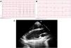
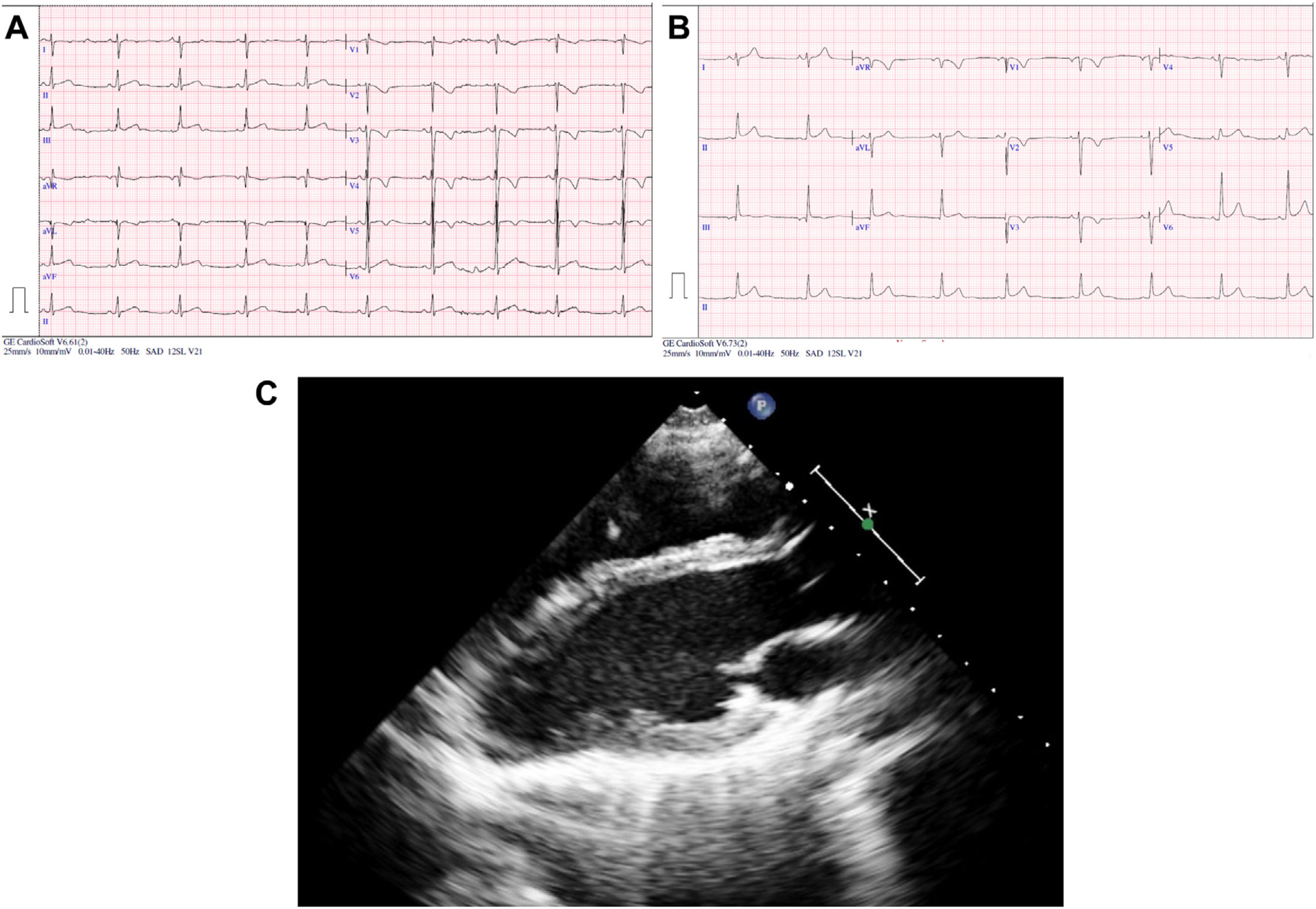
We present 2 cases of incidental finding of AoP. The first corresponded to a patient referred at the age of 13 years due to abnormal position of the heart in the chest radiography (CXR). He was symptomatic and did not have any relevant personal or family history. The physical examination was normal. The electrocardiogram (ECG) (Fig. 1A) showed sinus bradycardia with right axis deviation (+112 °), incomplete right bundle branch block (IRBBB), slow R wave progression, nonspecific intraventricular conduction delay (QRS duration of 96 ms) and abnormal repolarization (inverted T waves in V1–V4, transitioning in V5 and positive in V6). The CXR showed lung interposition between the aorta and pulmonary artery (PA), leftward shift of the heart with no visible right heart border and lung interposition between the left diaphragm and the base of the left heart (Fig. 2A–B). After performing an echocardiogram, PAoP was suspected on account of the posterior and leftward shift of the apex, paradoxical septal motion and apparent dilatation of the right ventricle due to leftward cardiac displacement (Fig. 1C; Supplemental material Appendix B, videos 1 and 2). Computed tomography (CT) and magnetic resonance imaging (MRI) findings confirmed the diagnosis (Fig. 2C–E). His exercise test was normal. As the patient was asymptomatic, a conservative approach was chosen. He has remained asmptomatic with no complications after 7 years follow-up.

Figure 1.

(A) Resting 12-lead ECG, patient 1. (B) Resting 12-lead ECG, patient 2. (C) Echocardiogram, patient 1. Long axis view showing posterior and rightward deviation of the apex of the left ventricle.

(0.61MB).
Figure 2.

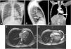
(A–C) Chest radiography, posteroanterior (A) and lateral (B); cardiac CT (C); leftward shift of the heart with lung interposition between the aorta and the pulmonary artery (*) and interposition of lung parenchyma between the left diaphragm and the border of the base of the left heart (arrow). (D) Cardiac MRI with the patient in the right lateral decubitus position. (E) Cardiac MRI with patient in supine position showing posterior shift of the cardiac silhouette (arrow).

(0.5MB).

The second patient was referred at the age of 5 years due to deviation of the heart in the CXR. During surveillance, she was diagnosed with Behcet disease. She was asymptomatic from the cardiac point of view and she did not have any other relevant personal or family history. She had normal physical examination. The ECG (at the age of 15 years, Fig. 1B) revealed mild right axis deviation (+94°), IRBBB, poor R wave progression, nonspecific intraventricular conduction delay (QRS duration of 98 ms) and abnormal repolarization (inverted T waves in V1–V3, transitioning in V4 and positive in V5–V6). The CXR and echocardiogram (supplementary material Appendix B, videos 3 and 4) revealed the same features described in patient 1. Her diagnosis was confirmed by MRI. She had a normal exercise test. A conservative management was chosen in view of the absence of symptoms. She has remained asymptomatic with no evidence of complications after 13 years of follow-up.

Although AoP is a rare disease, it is important to keep a high level of suspicion, mainly because patients with PAoP, in addition to being symptomatic more frequently, are at risk of herniation and strangulation of the left ventricle through the pericardial defect and, therefore, of sudden death.2,3,5

CXR, ECG, echocardiogram and CT are useful for diagnosis of AoP. However, the gold standard is MRI, which in black blood fast spin echo and steady-state free precession sequences evinces cardiac indentation at the location of the defect, interposition of lung tissue between the aorta and the PA or between the diaphragm and the base of the heart and leftward and posterior displacement of the heart. A rotation angle greater than 60° between the anterior-posterior line passing through the vertebral body and left ventricle main axis in the end-diastolic phase of the axial cine stack offers a good sensitivity for detection of left ventricular AoP.6

Although there is no global consensus on the management of these patients, close follow-up and monitoring for complications is recommended in asymptomatic patients in whom AoP is an incidental finding and in the case of complete bilateral or left-sided AoP. Surgery is reserved for patients with PAoP or at risk of complications.2,5

In conclusion, we have presented 2 cases of incidental diagnosis of AoP, a disease that is difficult to diagnose due to its rarity and usually asymptomatic, whose early detection is vital due to its potential association with sudden death. Although MRI is the gold standard of diagnosis, the abovementioned imaging investigations can also be useful. Despite the absence of a global consensus, the approach to treatment is conservative, reserving surgery for patients with symptoms or with PAoP at risk of complications.

Funding

This research did not receive any external funding.

Conflicts of interest

The authors have no conflicts of interest to declare.

Appendix A
Supplementary data

The following is Supplementary data to this article:

Bibliografía
[1]
M. Bouchard, A. Hoschtitzky, M. Gatzoulis.
Diagnosis and management of congenital absence of pericardium: a case report.
Eur Hear J Case Rep, 3 (2019), pp. 1-5
[2]
A.B. Shah, I. Kronzon.
Congenital defects of the pericardium: a review.
Eur Heart J Cardiovasc Imaging., 16 (2015), pp. 821-827
[3]
F. Verde, P.T. Johnson, S. Jha, E.K. Fishman, S.L. Zimmerman.
Congenital absence of the pericardium and its mimics.
J Cardiovasc Comput Tomogr., 7 (2013), pp. 11-17
[4]
B. Maisch, P.M. Seferović, A.D. Ristić, R. Erbel, R. Rienmüller, Y. Adler, et al.
Guidelines on the diagnosis and management of pericardial diseases: executive summary. The task force on the diagnosis and management of pericardial diseases of the European Society of Cardiology.
Eur Heart J., 25 (2004), pp. 587-610
[5]
A. Pérez-Casares, S. Cesar, L. Brunet-Garcia, J. Sanchez-de-Toledo.
Echocardiographic evaluation of pericardial effusion and cardiac tamponade.
Front Pediatr., 5 (2017), pp. 1-10
[6]
F. Macaione, A. Barison, I. Pescetelli, F. Pali, F. Pizzino, A. Terrizzi, et al.
Quantitative criteria for the diagnosis of the congenital absence of pericardium by cardiac magnetic resonance.
Eur J Radiol., 85 (2016), pp. 616-624

Please cite this article as: Brunet-Garcia L, Carretero Bellon JM, Pérez-Casares A, Prada Martínez FH, Sánchez-de-Toledo J, Ausencia parcial de pericardio: ¿únicamente un hallazgo incidental? Anales de Pediatría. 2022;96:540–542.

Copyright © 2021. Asociación Española de Pediatría
Download PDF
Idiomas
Anales de Pediatría (English Edition)
Article options
Tools
Supplemental materials