Journal Information
Vol. 98. Issue 3.
Pages 232-233 (1 March 2023)
Vol. 98. Issue 3.
Pages 232-233 (1 March 2023)
Images in Paediatrics
Open Access
Newborn inguinal hernia containing uterus, ovary and Fallopian tube
Hernia inguinal neonatal conteniendo útero, ovario y trompa de Falopio
Visits
2603
Javier Arredondo Monteroa,
Corresponding author
, Pilar Guillén Redondoa, Giuseppa Antonaa, Mónica Bronte Anautb
a Department of Paediatric Surgery, Hospital Universitario de Navarra, Pamplona, Spain
b Department of Pathology, Hospital Universitario de Araba, Vitoria, Spain
This item has received

Under a Creative Commons license
Article information
Full Text
Bibliography
Download PDF
Statistics
Figures (2)
Full Text

Indirect inguinal hernia is frequent in the paediatric population, being particularly prevalent in preterm infants1. In boys, it results from the persistence of the processus vaginalis, while in girls it results from the persistence of the canal of Nuck, an embryonic evagination of the parietal peritoneum that extends together with the round ligament through the inguinal canal until it reaches the labia majora. The most frequent content is intestinal loops, although in girls it may include, as in the case presented here, the ovary, the Fallopian tube and even the uterus2.

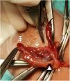
A 16-day-old newborn presented in our hospital with a mass in the left inguinal region of 48 hours of evolution. The patient was asymptomatic. Physical examination revealed the presence of an irreducible hernia in the left inguinal region with no associated cutaneous inflammation. An ultrasound scan evinced the presence of the ovary in the hernia, without vascular involvement (Fig. 1). An exploratory inguinotomy was performed, confirming the existence of an indirect inguinal hernia. The hernia contained the ovary, ovarian ligament, Fallopian tube, fimbriae and uterus (Fig. 2). The contents were reduced to the abdominal cavity and an herniorrhaphy was performed. The patient evolved favourably in the postoperative period. She is currently asymptomatic and under follow-up.

Figure 1.

A. Inguinal ultrasound. A hypoechoic mass measuring 9 × 14 mm with millimetric hypoechoic foci inside is identified within the hernial sac. B. Doppler ultrasound scan of the lesion which rules out vascular involvement.

(0.14MB).
Figure 2.

Intraoperative photograph. The ovary (white asterisk), ovarian ligament (white arrow head), Fallopian tube (solid white arrow), fimbriae (triple arrowhead) and uterus (dotted white arrow) were contained in the hernial sac, which is referenced with haemostat forceps.

(0.14MB).

When an inguinal hernia is surgically repaired in female patients, especially in newborns, it is important to consider that the hernia may contain an ovary. Although the presence of the uterus is extremely infrequent, with only 73 cases reported in the literature to date3, it is a possibility that must also be taken into account. A careful and methodical surgical examination prevents iatrogenic injuries.

References
[1]
V. Ramachandran, C.F. Edwards, D.C. Bichianu.
Inguinal hernia in premature infants.
Neoreviews, 21 (2020), pp. e392-e403
[2]
K. Karadeniz Cerit, R. Ergelen, E. Colak, T.E. Dagli.
Inguinal hernia containing uterus, fallopian tube, and ovary in a premature newborn.
Case Rep Pediatr, 2015 (2015), pp. 807309
[3]
G. Brooks, S. Bisoffi, C. Virgone, P. Gamba.
Inguinal hernia containing the uterus in the pediatric patient: A systematic review.
J Pediatr Adolesc Gynecol, 35 (2022), pp. 182-187
Copyright © 2022. Asociación Española de Pediatría
Download PDF
Idiomas
Anales de Pediatría (English Edition)
Article options
Tools
es en

¿Es usted profesional sanitario apto para prescribir o dispensar medicamentos?

Are you a health professional able to prescribe or dispense drugs?