


Intravascular devices are essential for the diagnostic and therapeutic approach to multiple diseases in paediatrics, and central venous catheters (CVCs) are especially important. One of the most frequent complications is the infection of these devices, which is associated with a high morbidity and mortality. These infections are highly complex, requiring the use of substantial resources, both for their diagnosis and treatment, and affect vulnerable paediatric patients admitted to high-complexity units more frequently. There is less evidence on their management in paediatric patients compared to adults, and no consensus documents on the subject have been published in Spain. The objective of this document, developed jointly by the Spanish Society of Paediatric Infectious Diseases (SEIP) and the Spanish Society of Paediatric Intensive Care (SECIP), is to provide consensus recommendations based on the greatest degree of evidence available to optimize the diagnosis and treatment of catheter-related bloodstream infections (CRBSIs). This document focuses on non-neonatal paediatric patients with CRBSIs and does not address the prevention of these infections.
Los dispositivos intravasculares son esenciales para el abordaje diagnóstico y terapéutico de múltiples patologías en pediatría, siendo especialmente importantes los catéteres venosos centrales (CVC). Una de las complicaciones más frecuentes es la infección de estos dispositivos, lo cual conlleva una elevada morbi-mortalidad. Estas infecciones presentan una gran complejidad, precisando de un elevado consumo de recursos, tanto para su diagnóstico como para su tratamiento, afectando de manera más frecuente a pacientes pediátricos vulnerables ingresados en unidades de alta complejidad. La evidencia para su abordaje en pediatría es menor que en adultos y no hay documentos de consenso realizados en nuestro medio. El objetivo de este documento, realizado entre la Sociedad Española de Enfermedades Infecciosas Pediátricas (SEIP) y la Sociedad Española de Cuidados Intensivos Pediátricos (SECIP), es dar recomendaciones de consenso basadas en la mayor evidencia disponible para optimizar el diagnóstico y tratamiento de las bacteriemias y fungemias relacionadas con el catéter. Este documento se centrará en pacientes pediátricos no neonatales, sin entrar en discusión sobre la prevención de estas infecciones.
Intravascular catheters are indispensable for the diagnosis and management of many diseases in paediatric care,1 and many children carry a central venous catheter (CVC), especially in complex care settings.
Infections related to CVCs is a frequent complication in paediatric patients, with a mean incidence of 0.9–3.5 infections/1000 catheter days,2–4 and it is the most frequent type of health care-associated infection in paediatric care.4 However, there is substantial heterogeneity among paediatric studies due to differences in the underlying diseases, types of catheters and applied terms and definitions. Many studies use the construct of “central line-associated bloodstream infection” (CLABSI) while others use the term “catheter-related bloodstream infection” (CRBSI). For the purposes of epidemiological surveillance, CLABSI is a simpler definition, as it does not require confirmation of infection of the CVC (Section 1 and Table 1). In this document we will refer to both as CRBSI.
Definitions specific to catheter-related bacteraemia/fungaemia.
| Concept | Definition | Comments |
|---|---|---|
| Central line-associated bloodstream infection (CLABSI) | A laboratory-confirmed bloodstream infection where an eligible organism is identified in a patient with a CVC when no other source of infection can be found | Used for epidemiological surveillance purposes, but overestimates infection of CVCs. |
| The CVC may not be the source of infection | ||
| Catheter-related bloodstream infection (CRBSI)a | Confirmed bloodstream infection whose source is the CVC | Requires confirmation of the catheter as the source of infection |
| Complicated CRBSIb | Catheter-related bloodstream infection with a poorer prognosis | Suppurative thrombophlebitisc, mycotic aneurism, disseminated infection (endocarditis, osteomyelitis, distant abscess), local complication (PAC abscess), septic shock and/or bacteraemia after ≥ 72 h of treatment |
| Blood culture contamination | Growth of a microorganism in a blood culture that is not really in the bloodstream and that was accidentally introduced in the specimen during the collection process | In paediatric care, it is usually harder to diagnose as more than 1 sample is not usually obtained in a single venepuncture procedure |
| Catheter colonization | Isolation of a microorganism in a CVC-drawn or catheter-tip blood culture in absence of isolation in venepuncture-drawn cultures and without suggestive clinical manifestations | Under certain circumstances, it may increase the risk of subsequent bloodstream infection |
| Local infection | Infection involving the CVC in the absence of systemic symptoms or bacteraemia/fungaemia. | Classified as: exit site infection (< 2 cm), tunnel infection (> 2 cm) and pocket infection |
CLABSI, central-line-associated bloodstream infection; CRBSI, catheter-related bloodstream infection; CVC, central venous catheter; PAC, port a cath.
See the section: When and how should blood cultures be done? Methods fo diagnosis of CRBSI and Fig. 1.
Although CRBSI is frequent in paediatric patients, high-quality evidence on the subject is scarce, which results in substantial heterogeneity in its management. Given the absence of consensus documents in paediatrics, we reviewed the available scientific evidence and developed recommendations to facilitate the diagnosis and treatment of this condition in the paediatric population.
This document applies to patients aged 1 month to 18 years with CRBSI, excluding patients in neonatal units or patients with infections associated with peripheral lines or other endovascular devices. It also does not address the prevention of CRBSI.
MethodsThis document has been developed with the collaboration of the Sociedad Española de Infectología Pediátrica (SEIP, Spanish Society of Paediatric Infectious Diseases) and the Sociedad Española de Cuidados Intensivos Pediátricos (SECIP, Spanish Society of Paediatric Intensive Care), with the participation of 7 specialists from these two societies and through the formulation of 11 questions. The scientific evidence was reviewed using the PICO model (patient [P], intervention [I], comparison [C] and outcome [O]). The quality of evidence and the strength of recommendation was assessed applying the grading system of the United States Department of Health and the guidelines of the European Society of Clinical Microbiology and Infectious Diseases (ESCMID) (Table 1S of the Supplementary data),1 and when the evidence was scarce, with the Delphi method. Once the document had been developed, 2 external evaluators and the corresponding SEIP/SECIP working groups revised it. The final recommendations can be found in Table 2.
Summary of the consensus recommendations for the management of central venous catheter-related bloodstream infections.
| What are the microorganisms most frequently involved in CRBSIs? (Table 3) |
| - The most frequent causative agents of CRBSIs in the paediatric population are gram-negative bacilli (GNB), chiefly, Enterobacterales and especially Klebsiella spp., and gram-positive cocci (GPC), especially Staphylococcus aureus and coagulase-negative Staphylococcus (CoNS) (A II) |
| - In contrast to the adult population, GNB are a frequent cause of CRBSI in children. (A II) |
| - Candida spp. are an infrequent cause of CRBSI.3,8,11 (A II) |
| When should a CRBSI be suspected? |
| - The presence of CRBSI should be ruled out in any child with a CVC who develops manifestations compatible with bacteraemia/fungaemia (especially in cases of fever without source or severe illness with organ dysfunction or septic embolism) (A II). Recent CVC use increases the risk of CRBSI, especially if catheter manipulation was difficult. |
| - If CRBSI is suspected, examine the CVC insertion site and its trajectory to rule out local infection. (A II) |
| - The presence of other clinical manifestations (respiratory, gastrointestinal) does not always rule out a CRBSI. (B II) |
| What are the risk factors associated with CRBSI in the paediatric age group? |
| - Underlying disease: children who undergo chemotherapy, haematopoietic stem cell transplantation, neutropenia, on total parenteral nutrition or tube feeding.11 (B II) |
| - Type of catheter: increased risk in temporary CVC and peripherally inserted CVCs; lower risk in tunnelled catheters and implanted catheters (lower risk with PAC).2,40 (B II) |
| - Increasing multilumen catheters number.4,7,12 Insertion of more than one CVC in place.7,12 (B II) |
| - Location of CVC. A few paediatric studies suggest an increased risk in CVCs inserted in the femoral vein.12 (C III) |
| - Catheter days in percutaneous catheters (>7 days).7 (B II) |
| - Previous history of bacteraemia.3,40 (B II) |
| When should blood cultures be performed?1,5,13 |
| - In every paediatric patient with a CVC and suspected CRBSI, blood cultures and/or CVC cultures should be performed, as appropriate (see below) before initiation of antibiotherapy. (A II) |
| - Culture of removed CVCs is not recommended in patients in whom CRBSI is not suspected. (A II) |
| How should samples for blood culture be taken? Criteria for CRBSI |
| - Whenever possible, a sample should be drawn from the CVC and another through venepuncture. (A II) |
| - In the case of CVCs with several lumens, a sample for culture should be drawn from each lumen. (A III) |
| - Due to the frequency of difficulties in venepuncture in paediatric patients, if obtaining a sample for blood culture through venepuncture is difficult, consider collection of samples from all catheter lumens and proceed as if one of the samples had been obtained by venepuncture. (B III) |
| - If the catheter is NOT REMOVED, the differential time to positivity (> 120 min) is the most appropriate technique, although it has limitations in terms of sensibility and specificity, especially for certain microorganisms (S. aureus and Candida spp.) (B II). There is little evidence on the use of this technique in CRBSIs caused by Candida spp. |
| - If the catheter is removed, submit the tip (last 5 cm) to the department of microbiology for culture (A II). For PAC ports, the entire PAC device should be submitted (reservoir and tip) for culture (BII). The most reliable catheter culture diagnostic methods are semiquantitative culture (roll plate or Maki technique) and quantitative culture (with vortexing or sonication) (A II). |
| What other diagnostic methods could be useful for CRBSIs? |
| - There is insufficient evidence to recommend routine use of molecular techniques for diagnosis of CRBSIs in paediatric clinical practice. (C III) |
| - Negative results in CVC surface cultures could help rule out CRBSI, given their high negative predictive value in adults. (B II) |
| - In cases in which suppuration is detected at the insertion site, in addition to blood specimens for blood culture, a sample of the exudate around the catheter exit should be collected for culture and Gram staining. (A III) |
| - Testing for biomarkers (eg, procalcitonin) could be useful when considering initiation of empirical antibiotherapy and to assess the clinical response, but there are no biomarkers specific for CRBSI. (B III) |
| When should additional testing be performed? |
| - Ruling out infective endocarditis (IE) by means of transthoracic echocardiography (TTE) is recommended in CRBSIs involving S. aureus in patients with: bacteraemia persisting for > 48 h of appropriate antibiotherapy, haemodialysis, metastatic foci, signs of IE, intravascular or intracardiac devices, prosthetic valves, previous history of IE or structural heart disease (A II). Individualize the use of TTE under any other circumstances (B II), and in case of microorganisms associated with a low risk of IE (CoNS, GNB) (B III). |
| - We suggest performance of echocardiography in patients with CRBSI caused by Enterococcus faecalis who experience seizures or neurologic complications or recurrence of bacteraemia, considering the presence of risk factors in all other patients. The risk of IE associated with other Enterococcus spp. is lower (B III). |
| - We recommend ruling out IE in patients with a CVC and candidaemia (A II). In addition, the diagnostic evaluation should also include a fundoscopic examination and the assessment of vascular thrombosis around the CVC (B II). |
| - Performance of trans-oesophageal echocardiography (TOE) should be considered in patients in whom visibility in the TTE can be limited (e.g., due to surgery or previous thoracic trauma), when intracardiac complications are suspected, or there is prosthetic material in place. (B II) |
| Which is the most appropriate antibiotic for empirical therapy? Should it be adapted based on the previous history of colonization/isolation? (Table 4) |
| - When CRBSI is suspected, and after collection of appropriate biological samples, empirical antibiotherapy should be initiated as soon as possible with an agent offering adequate coverage against GPC and GNB (A II). In patients without neutropenia, immunosuppression or extensive burn wounds who are clinically stable and have no underlying disease that could decompensate rapidly, not colonized by MDR organisms and in hospitals with a prevalence of multiple drug resistance < 5%–10% (e.g., methicillin-resistant S. aureus [MRSA], MDR GNB), empirical therapy could consist of cloxacillin (or cefazolin) + ceftriaxone (B III). Vancomycin (or teicoplanin) could be alternatives to cloxacillin, especially in settings with a high prevalence of MRSA. (B II) |
| - If the patient has risk factors for infection by Pseudomonas spp., such as neutropenia and other types of severe immunosuppression or extensive burns, use an antibiotic with a spectrum that covers this bacterium (A III). Consider the use of high doses and prolonged infusion. |
| - If there are local signs of infection or the symptoms start after recent difficulties in catheter handling, use antibiotics offering adequate coverage against CoNS and MRSA, such as glycopeptides (A II), or, as an alternative, if there is no pulmonary involvement, daptomycin (B III). Linezolid could be used if there is no suitable alternative (B III). |
| - In severe or potentially severe forms of disease (shock, multiple organ failure or patients with underlying disease that can decompensate rapidly) start broad-spectrum antibiotherapy including a glycopeptide or daptomycin (if there is no pulmonary involvement) combined one or two antibiotics against GNB (piperacillin-tazobactam, cefepime or meropenem/imipenem, and, depending on the risk of MDR and clinical severity, an aminoglycoside or fluoroquinolone). (B II) |
| - Children colonized by MDR organisms may be at increased risk of CRBSI caused by these organisms, and empiric antibiotherapy covering these organisms should be considered, especially in patients with severe illness and recent colonization (B II). Consultation with the ASP team is recommended. |
| - In the case of suspected CRBSI, admission of the patient with initiation of empirical antibiotherapy is recommended for a minimum of 24−48 h (A II). In specific situations in children with CVC and fever with no risk factors for severe disease and in good clinical condition, discharge can be considered with follow-up in 24 h after administration of a dose of antibiotics (for instance, ceftriaxone), or, alternatively, inpatient observation without antibiotherapy (especially if an alternative diagnosis is possible). (C III) |
| - In patients with β-lactam allergy, administer a glycopeptide (or daptomycin or linezolid) + aztreonam or a fluoroquinolone (B III). Meropenem and aztreonam have a very low cross-reactivity with penicillins (<1%) and could be administered safely in most patients (A II). Consultation with an allergist is recommended. |
| Antifungals for empirical treatment: When and which? |
| - Consider coverage of Candida spp. in empirical therapy in the case of suspected CRBSI in patients with several risk factors for this aetiological agent, especially in those with severe disease: femoral CVC, parenteral nutrition, prolonged antibiotherapy (especially with activity against anaerobic bacteria or vancomycin), immunosuppression, previous infection and/or colonization by Candida spp. at multiple sites. (B II) |
| - Based on the evidence in adults, in the case of a T2RM positive for Candida, coverage with an antifungal is recommended (B III). It should also be considered in the case of a positive result for β-D-glucan. (C III) |
| - The empirical treatment of choice is an echinocandin or liposomal amphotericin B (A I). Any of these options should also be used if it is not possible to remove the CVC on account of their activity against biofilm (in this case, liposomal amphotericin B at a dose of 3−5 mg/kg) (B III). |
| Which is the most appropriate directed antibiotic treatment? (Table 5) How should infections caused by multidrug-resistant organisms be treated? |
| S. aureus |
| - Cefazolin or cloxacillin are the treatment of choice in CRBSI caused by methicillin-sensitive S. aureus (MSSA). (A II) |
| - Vancomycin is the treatment of choice for CRBSI caused by MRSA (B II). Daptomycin (as long as there is no pulmonary involvement) would be the most adequate alternative (A II). Teicoplanin, ceftaroline or ceftobiprole can also be considered (B III). Linezolid could be considered in case of treatment failure or adverse events with other antibiotics. (C III) |
| - The daptomycin + ceftaroline combination is the most widely accepted treatment in the case of persistent CRBSI caused by MRSA despite adequate control of the source of infection (C III). |
| Coagulase-negative Staphylococcus |
| - We recommend initiation of cefazolin or cloxacillin in CRBSIs caused by methicillin-sensitive CoNS. (B II) |
| - In the case of methicillin resistance, vancomycin is the preferred option (B II), with daptomycin (if there is no pulmonary involvement), teicoplanin (if the pathogen is sensitive) or ceftaroline as possible alternatives. (B III) |
| Enterococcus spp. |
| - Ampicillin is the treatment of choice in CRBSIs caused by susceptible Enterococcus spp. (A II) |
| - Vancomycin is the treatment of choice in CRBSIs caused by Enterococcus spp. resistant to ampicillin. (B III) |
| - Linezolid or daptomycin are alternative options in case of vancomycin resistance. (B III) |
| Gram-negative bacilli |
| - Use the antimicrobials with the highest bactericidal activity, greatest safety and narrowest spectrum covering the isolated microorganism whenever possible. In this regard, β-lactam antibiotics are the preferred choice. (A II) |
| - In the case of MDR infection, consultation with a paediatric infectious disease expert is recommended to optimise treatment, given the risk of clinical worsening and complications. (B II) |
| When should the catheter be removed? |
| - In patients with severe local complications (tunnel infection, abscess associated with PAC) or infection of the insertion site associated with signs of systemic infection, or signs of persistent local infection despite 48−72 h of appropriate systemic antibiotherapy. (A II) |
| - Suppurative thrombophlebitis. (A II) |
| - Septic metastatic complications (endocarditis, osteomyelitis, pulmonary embolism), severe sepsis or septic shock. (A II) |
| - Persistent isolation of the same microorganism in blood cultures or persistent manifestations of bacteraemia after 48−72 h of appropriate systemic antibiotherapy (A II). |
| - Infection by S. aureus (A II), nonfermenting gram-negative bacilli (P. aeruginosa, Acinetobacter spp., Stenotrophomonas spp.), Candida spp. (A II), MDR microorganisms mycobacteria(B II), if an alternative source is not identified. |
| - In infections by Enterococcus spp., adult guidelines tend to recommend removal (although not in every case). Maintenance of the CVC could be considered in children. (B II) |
| - Infection by microorganisms that could cause IE (Staphylococcus spp. or Enterococcus spp.) in patients who carry intravascular devices. (A II) |
| - Consider in infections caused by microorganisms that are less virulent but difficult to eradicate (Bacillus spp., Micrococcus spp., Corynebacterium spp. Propionibacterium spp.). It is important to rule out contamination based on positivity of at least 2 cultures of CVC samples drawn at different times or from 2 different lumens or a CVC-drawn sample and an additional peripheral blood sample. (B II) |
| - If it is not possible to use antibiotic lock therapy and the CVC is the sole vascular access or the only available lumen and it is necessary for the patient management. (B II) |
| How should an exit site infection be treated? |
| - Tunnelled or haemodialysis catheters: in the case of exit site infection with no signs of systemic infection, tunnel infection or purulent drainage around the insertion site, and with negative culture results, consider initiation of topical antibiotherapy with mupirocin or a similar antibiotic (for infections caused by S. aureus) or clotrimazole (for infections caused by Candida spp.). If the infection persists beyond 48−72 h, consider systemic therapy with the possible addition of lock therapy before removing the catheter. (B III) |
| - Nontunnelled catheters (temporary CVC, peripherally inserted central catheter [PICC] or catheters with reservoirs like PAC): always remove, even in absence of signs of systemic infection (B II). However, given the potential difficulty of establishing vascular access in paediatric patients, conservative treatment without CVC removal could be considered in the case of permanent catheters. (C III) |
| When and how should a new catheter be inserted? |
| - The consensus group recommends the following (B II): |
| • Whenever possible, choose an insertion site different than the one used with the last CVC. |
| • Wait 24−48 h after the removal of the previous catheter, although in cases in which a catheter is absolutely necessary, it can be inserted after 12 h. In patients with candidaemia, it is better to wait 5 days. |
| • It would be advisable to wait to have a blood culture without growth (A MUST in CRBSIs caused by S. aureus and Candida spp.). |
| • Catheter replacement by guidewire exchange is generally not recommended. It can be done if there is no other available vascular access site or there is a high risk of bleeding diathesis. In such cases, we recommend use of a catheter impregnated with antibiotic and lock therapy for 8–12 h after recatheterization. (B III) |
| When is lock therapy indicated? |
| - Antimicrobial lock therapy may be considered in clinically stable patients in whom it would be difficult to insert a new CVC, who do not meet the criteria for catheter removal. This particularly applies to long-term catheters (B III). It must be taken into account that locking is less effective in temporary catheters, in which extraluminal colonization is more frequent. |
| - Under very specific circumstances in which placement of a different central catheter would be extremely difficult, lock therapy could be considered in infections caused by S. aureus or Candida spp. (C III) |
| - The antimicrobials used for lock therapy should have optimal activity against biofilm. Daptomycin, echinocandins and liposomal amphotericin B have exhibited good activity in vitro. (B II) |
| - This panel does not currently recommend locking with ethanol solution, given the low quality of the studies published to date, insufficient evidence on its effectiveness and reporting of multiple adverse events. (B III) |
| - Systemic antibiotherapy should always be administered simultaneously in patients with CRBSI, even if the CVC is treated with lock therapy. (A I) |
| Which are the optimal duration of treatment and route of administration (Table 7)? |
| - The duration of intravenous antibiotherapy for CRBSIs caused by S. aureus or Candida spp. should be ≥ 14 days. For the rest of commonly involved bacteria, it should be ≥7 days (B II). A duration of 10 days can be considered for treatment of S. aureus in select patients with no risk factors and in good clinical condition. (B III) |
| - For S. aureus and Candida spp., the duration should be calculated considering the first day in which the blood culture is negative as day 1 of treatment (B II). For other frequently involved microorganisms, day 1 is the first day of effective treatment. (B II) |
| - If the catheter is not removed, systemic antibiotherapy should be accompanied by lock therapy for catheter salvage for 10−14 days (possibly fewer days for some bacteria, such as CoNS), and both should be maintained for the entire course of treatment. (B II) |
| - In patients with uncomplicated CRBSI, with a favourable course of disease and who undergo catheter removal, de-escalation to oral therapy can be considered using agents with a high bioavailability (fluconazole, fluoroquinolones, trimethoprim-sulfamethoxazole or linezolid). (B II) |
ASP, antimicrobial stewardship programme; CRBSI, catheter-related bloodstream infection; CVC, central venous catheter; MDR, multidrug-resistant; PAC, Port-A-Cath.
Table 1 presents the different definitions related to CRBSI.
A CVC is an intravascular catheter that terminates at the heart or in one of the great veins. Permanent (long-term) central lines are tunnelled catheters (including dialysis catheters) and implanted catheters (including ports, e.g. Port-A-Cath [PAC]). The rest are temporary catheters. The most frequently used CVCs are described in Table 2S of the Supplementary data.
Bloodstream infection is the presence of a microbial pathogen in the bloodstream. Laboratory-confirmed bloodstream infection is defined based on the identified organism and the probability of colonization.6
- 1)
Bloodstream infection by recognised microbial pathogens NOT included in the United States National Healthcare Safety Network (NHSN) common commensal list6: Identification of the pathogen in one or more blood specimens is sufficient.
- 2)
Bloodstream infection by organisms INCLUDED in the NHSN common commensal list: must meet the following criteria:
- 1.
Symptoms compatible with CRBSI.
- 2.
Not related to an infection at a different site.
- 3.
The same microorganism is identified by culture from 2 or more blood specimens drawn in 24 h (a) from 2 different sites [such as: (1) 2 venepuncture samples, (2) one venepuncture and 1 CVC sample, or (3) different lumen of a single or more than one CVC], or (b) at different times.
- 1.
A CVC is considered the source of a bactaeremia/fungaemia if it has been in place for more than 2 consecutive calendar days and is no longer eligible as a source from 24 h from its removal.6
2What are the microorganisms most frequently involved in CRBSIs? (Table 3)Despite the aetiological variability between different paediatric CRBSI case series, and contrary to the findings in adults, a significant proportion of CRBSIs in paediatric patients are caused by gram negative bacilli (GNB)2,3,7 and by gram positive cocci (GPC), whereas infections by Candida spp. remain infrequent.8 Coagulase-negative Staphylococcus (CoNS) are a frequent cause of CRBSI, but their isolation must be interpreted with caution, as these organisms can colonise CVCs and contaminate blood cultures.9 As is the case of other colonizing microorganisms (NHSN organism list), it requires performance of additional blood cultures for confirmation5.
Microorganisms isolated most frequently in CRBSIs/CLABSIs in the paediatric age group.a
| Microorganism | Estimated frequencyb |
|---|---|
| CoNS | 14%–49% |
| Staphylococcus aureus | 10%–15% |
| Enterococcus spp. | 4%–27% |
| Total gram-positive bacteria | 37%–66% |
| Enterobacterales | 16%–47% |
| Pseudomonas spp. and other nonfermenting gram-negative bacilli | 5%–21% |
| Total gram-negative bacilli | 27%–41% |
| Candida spp. | 3%–8%c |
| Polymicrobial | 7%–35% |
CLABSI, central-line-associated bloodstream infection; CoNS, coagulase-negative Staphylococcus; CRBSI, catheter-related bloodstream infection.
Fever, chills and gastrointestinal symptoms are the most frequently reported symptoms.10 Between 21% and 33% of patients with a CRBSI have respiratory symptoms, 20% hypotension, and fewer than 10% local manifestations.11 However, the symptoms may be nonspecific. It is important to rule out CRBSI when the manifestations are suggestive of disseminated infection (e.g. septic embolism) or CVC malfunction.1 It is important to always assess for other possible causes of the symptoms, such as community-acquired infections.
There is little evidence on the risk factors for CRBSI in paediatrics, which we summarise in Table 2, and Table 3S of the Supplementary data (Appendix).2,3,7,11,12
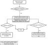
4When and how should blood cultures be done? Methods for diagnosis of CRBSI (Figs. 1 and 2)Collection of specimens for blood culture is recommended in patients with CVCs and manifestations compatible with CRBSI before initiation of antibiotherapy. Blood samples should not be drawn from central lines or through peripheral venepuncture in patients in whom CRBSI is not suspected.1,5,13


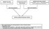
Different diagnostic methods can be used to confirm the involvement of a CVC in a CRBSI (Fig. 1), although none of them is completely reliable14:
- A.
Diagnosis of CRBSI without removal of the CVC. Blood specimens measuring the same volume should be collected at the same time from each catheter lumen and by venepuncture (at least 1 venepuncture specimen) for blood culture.1 When venepuncture is not an option, at least 2 specimens should be drawn from catheter lumens.5,15 The rationale for these diagnostic methods is that if the CVC is the source of the infection, a greater concentration of the causative pathogen will be inoculated in the corresponding cultures, with earlier detection of microbial growth (differential time-to-positivity [DTP]) or more marked growth (quantitative blood culture) compared to cultures of venepuncture samples.16
- -
Differential time-to-positivity cultures are used more frequently because they are simpler to perform. Detection at least 120 min earlier in the CVC-drawn culture compared to the venepuncture-drawn culture offers a high sensitivity and specificity for diagnosis of CRBSI,14,16 with a lower sensitivity for S. aureus and a lower specificity for Candida spp.16 It is preferable to draw the venepuncture specimens first, ensuring the same volume is used for each culture, at maximum 15−30 min earlier.
- -
Quantitative blood culture offers a high sensitivity and specificity for diagnosis of CRBSI, but requires more effort and resources.1
- -
As a general rule, it is very important to collect an adequate volume of blood per bottle14: >0.5–3 mL in infants under 1 year, 2–4 mL in children aged 1–3 years, 3–8 mL in children aged 5–10 years and 10 mL in those aged more than 10 years (weight > 20−30 kg). Some authors recommend larger volumes.14 Volumes of less than 10 mL should be inoculated in paediatric blood culture bottles and larger volumes in adult bottles.14 In the paediatric population, the incidence of bacteraemia by anaerobic organisms is less than 1%, therefore anaerobic blood culture bottles are not used except in specific cases, such as suspicion of intraabdominal infection, and prioritising the appropriate volume (in case of an insufficient volume, only the aerobic culture bottle would be inoculated).1,14Streptococcus pneumoniae and other gram-positive bacteria may also be isolated in anaerobic bottles.14
- B.
Diagnosis of CRBSI with removal of the CVC. In these cases, the catheter tip (5 cm) or the whole PAC should be submitted for culture.5,14 With this method, CRBSI is defined as isolation of the same microorganism in the cultures of venepuncture and catheter/reservoir specimens (Fig. 2):
- -
Semiquantitative culture: ≥ 15 UFC/plate 24 h after rolling the CVC segment over blood agar (Maki roll plate technique). This method retrieves microorganisms from the external surface of the CVC.14
- -
Quantitative culture. It is more arduous. An additional technique (flushing, sonication) is used to obtain intraluminal material which is then seeded in blood agar.5,13,14
- -
These techniques may be useful for diagnosis of CRBSI along with blood cultures, especially after initiation of antibiotherapy.1 They offer a quicker turnaround time, although they may be less sensitive than blood culture in paediatric patients.17 There are other disadvantages: a smaller spectrum of identifiable pathogens, limited antibiotic susceptibility testing, lesser availability and higher costs. Yet another drawback is the additional amount of blood that needs to be collected.
MALDITOF-MS (matrix-assisted laser desorption/ionization-time of flight mass spectometry)Reduces the time to identification in positive blood cultures and could allow early initiation of appropriate antibiothera.1,14
Culture of surface specimen from catheter insertion siteWhen CRBSI is suspected, culture of specimens obtained from the surface of hubs, the catheter surface at the insertion site and the skin surrounding the catheter offers a high negative predictive value in adults.1 Such samples should always be obtained from culture if there is suppuration/exudate at the site of insertion (Fig. 2).1,5
BiomarkersProcalcitonin offers a high sensitivity for detection of bacteraemia in children with indwelling CVCs and fever after excluding other sources of bacterial infection.18 In addition, it can be useful to monitor the patient’s response. The usefulness of other biomarkers has not been studied in depth. Fungal biomarkers are discussed in Section 8.
6Should additional diagnostic tests be used?EchocardiogramThe risk of infective endocarditis (IE) in patients with CRBSI depends on the aetiological agent and potential predisposing factors.1 Infective endocarditis is rare in children without cardiac disease, even in the case of bacteraemia caused by S. aureus, and a transthoracic echocardiogram should be performed in patients with symptoms suggestive of IE or relevant risk factors (Table 2).19,20 In other situations and in children with CRBSI involving pathogens unlikely to cause IE (CoNS, GNB), the decision should be individualised.1
Enterococcus faecalis is associated with IE in adults1,21 and, based on studies in adults, performance of echocardiography is recommended in patients with CRBSI by E. faecalis who develop neurologic complications or experience recurrence of bacteraemia (consult additional criteria applied in adults in the full text document). The risk of IE is much lower for other Enterococcus spp.
The risk of IE following candidaemia is of 1%–3%, but this condition is associated with a high morbidity and mortality22,23 and a high frequency of septic embolism, so performance of echocardiography is recommended.19,22,24 In patients with candidaemia, it is also necessary to perform a fundoscopic examination,23,24 an abdominal ultrasound examination and to rule out vascular thrombosis.
Transoesophageal echocardiography may be useful in cases with limited visibility, when intracardiac complications are suspected or when there is prosthetic material.19
7Which is the most appropriate antibiotic for empirical therapy? Should it be adapted based on the previous history of colonization/isolation?When CRBSI is suspected, antibiotherapy should be initiated early (within 60 min), as it improves outcomes.25Table 4 details the recommended empirical antibiotherapy for different clinical situations. Some experts recommend initiation of systemic antibiotherapy in any paediatric patient carrying a CVC with fever.11 However, in children without risk factors for severe CRBSI (Tables 2 and 4) in whom there have been no difficulties with catheter handling, without signs of local infection, without severe illness or an underlying disease that carries the risk of rapid decompensation, the initial approach can be close monitoring without antibiotherapy or administration of a parenteral antibiotic, such as ceftriaxone,26 while awaiting the results of blood cultures and other microbiological tests.
Empirical treatment for suspected CRBSIs.
| Condition at initiation | Microorganism | Antibiotherapy | Comments |
|---|---|---|---|
| Clinically stableNo underlying disease or risk factorsaNo high prevalence nor colonisation by MDR organisms | SASMbGNBb (except Pseudomonas spp.) | Cloxacillin/cefazoline+ ceftriaxone | In low-risk patients, consider:- Outpatient management with (ceftriaxone) and follow-up in 24 h- Observation without antibiotherapy, especially if another aetiology is possible |
| Severe immunosuppressionc or extensive burns | Pseudomonas spp. | Replace ceftriaxone by an antipseudomonal β-lactam agent. | There could be other risk factors, such as cystic fibrosis |
| Recent difficulty in catheter handling or local signs of CVC infection, high prevalence (> 5%–10%) of MRSA | MRSA, CoNS | Replace cloxacillin/cefazoline by a glycopeptided | |
| Colonization or previous infection by MDR organism | Several options (ESBL-, carbapenemase-producing organisms…) | Variablee | Consult with paediatric infectious disease specialist |
| Severe or potentially severe form of diseasef | MRSA, Pseudomonas spp. | Glycopeptided + antipseudomonal β-lactamg | Consider double coverage against GNB (adding fluoroquinolones or an aminoglycoside) |
| Risk of candidaemia | Echinocandin or liposomal amphotericin B | Consider initiation of an antifungal in the case of severe illness in children with several risk factorsh | |
| β-lactam allergyi | Glycopeptide/daptomycin + aztreonam/fluoroquinolone | Meropenem (<1% cross-reactivity with penicillin) |
CVC, central venous catheter; CRBSI, catheter-related bloodstream infection; ESBL, extended spectrum β-lactamase; GNB, gram-negative bacilli; MDR, multidrug-resistant; MRSA, methicillin-resistant S. aureus; MSSA, methicillin-sensitive Staphylococcus aureus.
Situations that can increase the risk of infection by other bacteria: colonization or previous infection by MDR organism, high prevalence of MDR organisms in the facility, previous history of bacteraemia, recent difficulty in catheter handling (24−48 h), severe immunosuppression,c extensive burn wounds, parenteral nutrition. Temporary CVCs, especially those dwelling for more than 7 days, carry a higher risk of CRBSI, so this section rather refers to long-term/permanent CVCs. Broader coverage could also be considered in patients with underlying disease that could decompensate rapidly.
Whenever CRBSI is suspected, antibiotherapy with adequate coverage against these bacteria should be initiated.
Vancomycin as first-line treatment. Alternative: daptomycin (in absence of pulmonary involvement) or linezolid.
Coverage for MDR organisms, especially in the case of severe illness or recent colonization (especially in the last 7 days). Consider the epidemiology of the unit/facility.
In patients with very severe infection (eg, septic shock) consider administration of a carbapenem (meropenem/imipenem).
Empirical antifungal therapy is rarely initiated in children with suspected CRBSI due to the low frequency of a fungal aetiology. Consider initiation in patients with several risk factors, especially those with severe forms of infection. Risk factors associated with candidaemia: femoral CVC, parenteral nutrition, antibiotherapy against anaerobic bacteria or vancomycin and a cancer diagnosis.31 Other risk situations in which empirical antifungal therapy should be considered are: prolonged stay in the paediatric intensive care unit, prolonged antibiotherapy, immunosuppression, gastrointestinal surgery, previous history of candidaemia without catheter removal and/or colonization by Candida spp. in several sites.
Adults colonized by multidrug-resistant (MDR) bacteria are at high risk of becoming infected by them, especially in cases of recent colonization (e.g., ≤ 7 days).27 Studies in children have also found an increase in this risk, with greater clinical severity,28 although it can probably be attributed to inadequate empirical therapy.29 Given the recent increase in bacteraemia caused by MDR organisms,3,7,30 it is advisable to consider the colonization status for empirical antibiotherapy, especially in severely ill patients. Consultation with antimicrobial stewardship teams is recommended in these situations.1
The recommended dosage for antibiotherapy can be found in https://www.seipweb.es/dosisantibioticos/. It is important to consider measurement of plasma drug levels as indicated in the summary of product characteristics, for instance in the case of treatment with glycopeptides o aminoglycosides.
8Antifungals for empirical treatment: when and which?Candida spp. are the most frequent fungal agents involved in CRBSIs in children,22 although must less frequently than in adults. Therefore, the indication of including an antifungal agent for empirical treatment of suspected CRBSI in paediatric patients should be individualised, considering the severity of disease and the presence of associated risk factors (Tables 2 and 4).22,31
The new rapid diagnostic tests for detection of invasive fungal infection can guide the decision to initiate antifungal treatment. In adults, β-d-glucan testing seems to be useful, but the evidence is insufficient in the paediatric age group. Although the T2 magnetic resonance assay technique for detection of candidiasis has not been studied specifically for diagnosis of CRBSIs, it may be useful in patients admitted to the Paediatric Intensive Care Unit (PICU) with suspected invasive candidiasis.32
The empirical treatment of choice for a CRBSI caused by Candida spp. is an equinocandin,1,24 as these drugs offer adequate anti-biofilm activity.1,33 High-dose liposomal amphotericin B is a good alternative that is also active against biofilm.1
9Which is the most appropriate directed antibiotic treatment? (Table 5). How to treat infections by multidrug-resistant organisms?S. aureusThe first-line treatment for methicillin-sensitive S. aureus (MSSA) is intravenous cefazolin/cloxacillin.1 In patients with β-lactam allergy or with methicillin-resistant S. aureus (MRSA), vancomycin or daptomycin may be used (if there is no pulmonary involvement).20 Vancomycin at a dose of 60 mg/kg/day achieves an AUC24/MIC (area under the serum concentration vs time curve for 0−24 h/minimum inhibitory concentration), greater than 400 in children, which is a better pharmacokinetic parameter than trough levels.20
Directed therapy recommended for treatment of CRBSIs.
| First-line | Alternativea | |
|---|---|---|
| MSSA | CefazolinCloxacillin | Vancomycin/teicoplaninDaptomycinb |
| MRSA | VancomycinDaptomycinb | TeicoplaninCeftaroline/ceftobiproleLinezolid |
| CoNSc | Vancomycin/teicoplanin | DaptomycinbCeftaroline |
| Enterococcus spp.d | Ampicillin | Teicoplanin/vancomycinLinezolidDaptomycinb |
| Escherichia coli and susceptible enterobacteria | Ampicillin | Cefotaxime/ceftriaxoneFluoroquinolones |
| Pseudomonas spp.*,e | Ceftazidime | Cefepime/piperacillin-tazobactam/aztreonamMeropenem/imipenemFluoroquinolonesNovel β-lactamsf |
| ESBL-producing Enterobacterales* | Ertapenem/meropenem/imipenemg | Piperacillin/tazobactamhNovel β-lactamsf |
| Enterobacterales with high probability of AmpC derepression*,i | Cefepime (if MIC ≤ 2 µg/mL) | Ertapenem/meropenem/imipenemgNovel β-lactamsf |
| Carbapenem-resistant bacteria* | Varies depending on resistance mechanismNovel β-lactamsf |
AmpC, class C β-lactamase (broad spectrum) ; CoNS, coagulase-negative Staphylococcus; ESBL, extended spectrum β-lactamase; MIC, minimum inhibitory concentration; MRSA, methicillin-resistant Staphylococcus aureus; MSSA: methicillin-sensitive S. aureus.
Depending on susceptibility, availability, allergic reactions, adverse effects. It must be taken into account that some of these agents are not authorised for this indication in paediatric patients, but they may be adequate alternatives based on the previous experience with their use in paediatric care and their known efficacy in adults or for treatment of other severe infections. Consultation of the summary of product characteristics is recommended.
Avoid in patients with pulmonary involvement, as it is inactivated by surfactant. It may be effective for pulmonary septic embolisms caused by distant sources, but caution should be exerted.
In the rare cases in which a CoNS isolate is susceptible to methicillin, treat like MSSA. Some strains are resistant to teicoplanin.
Use high doses of daptomycin (8−12 mg/kg/day) and teicoplanin (12 mg/kg/day) (https://www.seipweb.es/dosisantibioticos/).
In susceptible isolates, best as monotherapy38: ceftazidime-avibactam (authorised for paediatric use), ceftolozane-tazobactam (almost specific for treatment of Pseudomonas spp. that do not produce carbapenemases), cefiderocol.
In select cases in which it is initiated as empirical therapy and the patient exhibits improvement after the isolation is confirmed.38
In the case of MDR organisms, we recommend consultation with a paediatric infectious disease specialist. Recommended reference: Protocolo Resistencias Bacterianas en Pediatría (AEP 2023 bacterial drug resistance protocol for paediatric care): https://www.aeped.es/sites/default/files/documentos/2_resistencias_bacterianas.pdf.
In general, vancomycin/teicoplanin (there are strains resistant to teicoplanin1) are the treatment of choice, and daptomycin an adequate alternative34. Ceftaroline could be another option, although there is little evidence on its use in children with bacteraemia. The first-line treatment for methicillin-sensitive strains is cefazolin/cloxacillin.1S. lugdunensis should be managed like S. aureus.1
Enterococcus sppAmpicillin is the first-line treatment for CRBSIs caused by Enterococcus spp. in sensitive strains,1,5 and vancomycin is the treatment of choice for resistant strains.1 Linezolid or daptomycin (8−12 mg/kg/day) are possible alternatives,1,35 although there are no paediatric studies reporting their use in CRBSIs.34,36
Gram-negative bacilliDirected antibiotherapy for CRBSI caused by GNB should be based in the results of antimicrobial susceptibility testing, especially in the case of MDR organisms,1,35 and consultation with a specialist in paediatric infectious disease is recommended.1 Optimization of antibiotherapy against certain bacteria and in cases of severe infection is advisable: high doses and prolonged infusion35 (Pseudomonas spp., MDR) (https://www.seipweb.es/dosisantibioticos/).
10Central venous catheter management: when is removal or antimicrobial lock therapy indicated?A. When should the catheter be removed and how should it be handled? How should a local catheter infection be treated? When should another central line be inserted?To determine whether a CVC should be removed, it is important to take into account the type of catheter (temporary/permanent), the feasibility of inserting another one, the immune status/underlying disease of the patient and the infection.1,13
In general, immediate removal of the catheter after diagnosis of CRBSI is not necessary in haemodynamically stable patients who do not carry any other intravascular devices or, in the case of signs of local infection, without evidence of bacteraemia/fungaemia (Table 2).1,37,38 Higher rates of treatment failure have been reported when the CVC was not removed in CRBSIs caused by S. aureus,39P. aeruginosa, Enterococcus spp., Candida spp.,38 mycobacteria and MDR organisms.1,38 However, some paediatric studies have reported high success rates in patients with CRBSI caused by SAMS,39,40 although the existing evidence is not of high quality. A systematic review in children found high rates of successful catheter salvage with intravenous antibiotherapy with the addition of a lock solution in CRBSIs caused by MSSA, CoNS, GNB and Enterococcus spp., with a much lower rate for MRSA.37 This study found a higher rate of recurrence in CRBSIs caused by MRSA, in cases treated with vancomycin lock or in cases without lock treatment. Once the withdrawal of the CVC has been decided, it should be rapidly done (6–12 hours if clinical severity) according to another pediatric study.39
Catheter processing1- -
Disinfect the skin around the catheter with ethanol/chlorhexidine.
- -
Prevent air embolism in catheters inserted in the superior vena cava.
- -
Submit the catheter tip (5 cm) or the entire PAC in a sterile container (or store at 4–8 °C and submit as soon as possible) for culture.
The treatment of an exit-site infection depends on the type of catheter, and it can be more conservative for long-term catheters (Table 2). In the case of tunnel infection, the CVC should always be removed combined with administration of a 7–10 day course of systemic antibiotherapy (14 days for S. aureus).1,5
After removing the catheter, it is better to delay insertion of a new catheter for 24–48 h (5 days for Candida).1,10 Replacement of the CVC by guidewire exchange does not reduce the risk of new episodes of CRBSI.12 Thus, a prospective study in children admitted to the PICU with CVCs found an increased risk of CRBSI after replacement by guidewire for non-infectious reasons. If replacement were necessary, the use of antibiotic-impregnated CVCs would be advisable, as there is evidence of a decrease in CRBSIs with the prophylactic use of these CVCs in paediatric patients.41
B. When is antimicrobial lock therapy indicated?In the event of a CRBSI, catheter removal continues to be the preferred approach. However, when maintaining the catheter is critical, if removal is not absolutely necessary (Section 10.A and Table 2) one possibility is to maintain it by administering an antimicrobial lock solution in combination with systemic therapy (especially for permanent CVCs).37 Clinical trials in adults have found CVC locking in association with systemic antibiotherapy successful.1,5 A paediatric systematic review found a salvage success rate of 75%.37
The goal of lock therapy is to act on the biofilm, especially in long-term CVCs, in which there is a greater proportion of intraluminal colonization.33,38 It consists in filling the CVC (2−3 mL) with an anticoagulant solution with a high antibiotic concentration and not using that lumen for the duration of the therapy (the best option is a continuous lock, but, if not possible, the dwell time should be a minimum of 8−12 h/day), with maximum dwell times before lock replacement of 48–72 h.1,33,42 The solution should be prepared under sterile conditions, preferably in pharmacy settings.1,33 Before using the CVC or replacing the lock solution, the catheter should be aspirated and a volume equivalent to the administered lock volume removed.1,42
Until the infected lumen is confirmed, some experts recommend alternating the lumens to which the lock solution is administered and, if this is not possible, rotating the lumens through which intravenous antibiotherapy is administered.1,5,42
Two paediatric randomised trials of ethanol lock therapy with ethanol 70% have been published. One of them, which included a small number of patients with CRBSIs, compared two different ethanol locks,43 while the other found that treatment failure was similar in the ethanol lock and placebo groups, with a higher frequency of adverse events in the ethanol group.44 Therefore, we do not recommend initial locking with ethanol.
The antibiotics used for locking (Table 6) have to fulfil a number of criteria1,33,42:
- 1.
High activity against biofilm, such as daptomycin or echinocandins/amphotericin B.
- 2.
Concentrations much larger than the MIC.
- 3.
Stable at room temperature.
- 4.
Compatible with anticoagulants.
- 5.
Low resistance induction potential.
Solutions for antimicrobial lock-therapy.1,33,44
| Antibiotic group | Antibiotic | Concentrationa | Heparina | Comments |
|---|---|---|---|---|
| Cephalosporins | Cefazoline | 5−10 mg/mL | 100 U/mL | 2500 IU/mL in some protocols |
| Ceftazidime | 5 mg/mL | 2.500 U/mL | ||
| Aminoglycosides | Amikacin | 5 mg/mL | No | Amikacin 50 mg/1 mL; dissolve 1 mL in 9 mL physiological saline. |
| Amikacin | 1.5-2 mg/mL | 20 UI/mL | 1250 UI/mL in some protocols | |
| Gentamicin | 2−5 mg/mL | 20 UI/mL | Gentamicin 40 mg/2 mL; dissolve 2 mL in 6 mL de physiological saline | |
| Glycopeptides | Teicoplanin | 10 mg/mL | 2500 IU/mL | Preparation: dissolve teicoplanin in heparin |
| 2 mg/mL | 10 UI/ mL | |||
| Vancomycin | 2−5 mg/mL | 100 UI/mL | 2500 IU/mL in some protocols | |
| Fluoroquinolones | Ciprofloxacin | 2 mg/mL | No | Precipitates with heparin |
| Levofloxacin | 5 mg/dl | No | Precipitates with heparin | |
| Lipopeptide | Daptomycin | 3.5-5 mg/mL | 100−4.000 UI/mL | High dose in case of dialysis |
| Dilute in Ringer’s lactate solution | ||||
| Oxazolidinone | Linezolid | 2 mg/mL | 10 UI/mL | |
| Antifungals | Caspofungin | 2−3 mg/mL | No | |
| Micafungin | 1 mg/mL | 100 UI/mL | ||
| Amphotericin B | 2 mg/mL | No |
Ideally, it should be prepared in the pharmacy in single-dose containers.
IU, international units.
The duration of treatment depends on whether or not the CVC is removed and the potential complications of the CRBSI (Table 2),1 and treatment of complicated CRBSI should be individualised.
If the CVC is removed due to suspected sepsis, in absence of bacteraemia/fungaemia, but with colonization of the catheter by S. aureus, Candida spp. or nonfermenting GNB, intravenous antibiotherapy should last 3–5 days (5−7 days for S. aureus and up to 14 days for Candida).5 In case of infection by CoNS and fermenter GNB, additional treatment may not be necessary.13 The removal of a colonized CVC with no signs of infection does not require treatment, but it does require clinical surveillance with follow-up blood cultures for S. aureus, Candida spp. and Nonfermenting GNB.13
Duration of antibiotherapy for CRBSI based on the clinical presentation and causative agent.
| Type of infection or pathogen | Approach | |
|---|---|---|
| Complicated infection | Tunnel infection or implanted port abscess (long-term CVC) | • Remove catheter or port• Cleaning and drainage. Systemic antibiotherapy for at least 7−10 daysa |
| Systemic complications (e.g., septic thrombosis, endocarditis, osteomyelitis) | • Remove catheter or port• Systemic antibiotherapy 4−6 weeksa | |
| Uncomplicated infectionb | Staphylococcus coagulase-negatives | • CVCc removed: systemic antibiotherapy 5−7 days (or even 3 days) |
| • CVC salvaged: systemic antibiotherapy and lock therapy for 10−14 dayse | ||
| Staphylococcus aureusStaphylococcus lugdunensis | • Remove CVC: systemic antibiotherapy ≥ 14 daysf | |
| Enterococcus spp. | • CVC removed: systemic antibiotherapy 7−14 daysg | |
| • CVC salvage: systemic antibiotherapy 10−14 days and lock therapy for 10−14 days | ||
| Gram-negative bacilli | • CVC removedh: systemic antibiotherapy for ≥ 7 days (7−10 days in immunocompromised patients) | |
| • CVC salvaged: systemic antibiotherapy and lock therapy ≥ 10 days | ||
| Candida spp. | • Remove CVC: systemic antifungal ≥ 14 days | |
CRBSI, catheter-related bloodstream infection; CVC, central venous catheter.
In the case of S. aureus y Candida spp., day 1 of treatment corresponds to the first day in which the blood culture turns out negative. For other common microorganisms, it corresponds to the first day of effective treatment. If the CVC is removed and there is clinical improvement, blood cultures should be repeated in the case of CRBSI caused by S. aureus or Candida spp., but there is no evidence on the need to repeat blood cultures for infections by other microorganisms.1
Take into account that the response to lock therapy is better in patients with long-term CVCs, as there is a higher proportion of intraluminal colonization of these devices (in the case of CVC salvage). If the CVC is salvaged, follow-up blood cultures should be performed at 48–72 hours in all cases.
Therefore, if the CVC is not removed, specimens must be collected for blood culture 48–72 h after treatment initiation,1 whereas if the catheter is removed, performance of blood culture for follow-up is required for S. aureus and C. albicans, but not for other pathogens if there is clinical improvement.1
Sequential oral therapyRecent studies in adults suggests that after CVC removal and 3–9 days of intravenous treatment, switching to oral antibiotherapy seems safe and effective in uncomplicated CRBSI caused by gram-positive bacteria.1,45 In the case of CRBSI caused by Candida spp., in stable patients without metastatic complications who have undergone catheter removal, de-escalation to fluconazolee, even oral fluconazolee, can be contemplated.1,46
Thus, in patients with CRBSI that undergo CVC removal (or without CVC removal in infections by CoNS/GNB), without risk factors and with a favourable course of disease, de-escalation to oral therapy can be considered as long as the antimicrobial has a high oral bioavailability, for instance, with fluoroquinolones, trimethoprim-sulfamethoxazole, linezolid1 or fluconazolee.
Conflicts of interestJSL has collaborated as a speaker in an industry symposium sponsored by Biomerieux at the Annual Meeting of the European Bone and Joint Infection Society (EBJIS) in 2023. In 2023, he received funding from CIBERINFEC for the study of invasive disease by Streptococcus pyogenes and to attend the Annual Meeting of the European Society for Pediatric Infectious Diseases (ESPID). CG has received funding from the Spanish Ministry of Science and Innovation -Instituto de Salud Carlos III and the European Regional Development Fund (ERDF) (Juan Rodés grant JR22/00044). He has also received a research and education grant from Gilead and collaborated GSK, MSD and Pfizer clinical trials. All other authors have no conflicts of interest to disclose.
We thank María Guembe and Iolanda Jordán for their kind, disinterested and meticulous revision of the manuscript. We are also grateful for the collaboration and support of the working groups of the Sociedad de Infectología Pediátrica (SEIP) and the Sociedad de Cuidados Intensivos Pediátricos (SECIP), whose members we detail below:
The following is Supplementary data to this article:

Coordinator: Leticia Martínez Campos (Hospital Universitario Torrecárdenas, Almería). Secretary: Jesús Saavedra Lozano (Hospital Gregorio Marañón, Madrid). Members: Jaime Carrasco Colom (Hospital Universitario Son Espases, Majorca), Cristina Calvo Rey (Hospital la Paz, Madrid), Elena Colino (Complejo Hospitalario Insular MaternoInfantil Las Palmas de Gran Canaria), Ana Isabel Menasalvas Ruiz (Hospital Virgen de la Arrixaca, Murcia), Esmeralda Núñez Cuadros (Hospital Regional de Málaga, Malaga), Carlos Rodrigo Gonzalo de Liria (Hospital Germans Trias i Pujol, Badalona), David Aguilera Alonso (Hospital Gregorio Marañón, Madrid), David López (Hospital Costa del Sol, Marbella) and Elvira Cobo Vázquez (Hospital Universitario Fundación de Alcorcón, Madrid).
Coordinator: Luis Escosa (Hospital la Paz, Madrid). Members: Walter Alfredo Goycochea Valdivia (Hospital Virgen del Rocío, Seville), Ana Isabel Menasalvas Ruiz (Hospital Virgen de la Arrixaca, Murcia), Olaf Neth (Hospital Virgen del Rocío, Seville), Anabel Piqueras (Hospital la Fe, Valencia), María del Mar Santos Sebastián (Hospital Gregorio Marañón, Madrid), Eneritz Velasco (Hospital Sant Joan de Deu, Barcelona), David Aguilera Alonso (Hospital Gregorio Marañón, Madrid), Laura Martín Pedraz (Hospital Regional de Málaga, Malaga), Daniel Blázquez Gamero (Hospital 12 de Octubre, Madrid) and Ángela Manzanares (Hospital 12 de Octubre, Madrid).
Coordinator: Peter Olbrich (Hospital Virgen del Rocío, Seville). Secretary: Begoña Carazo Gallego (Hospital Regional Universitario de Málaga), Laura Ferreras Antolín (St George’s Hospital, London), Marisa Navarro Gómez (Hospital Gregorio Marañón, Madrid), Olaf Neth (Hospital Virgen del Rocío, Seville), Pere Soler Palacín (Hospital Vall d’Hebrón, Barcelona), José Tomás Ramos Amador (Hospital Clínico San Carlos, Madrid), Elena Rincón López (Hospital Gregorio Marañon, Madrid), Carlos Daniel Grasa Lozano (Hospital La Paz, Madrid), Natalia Mendoza (Hospital Vall d’Hebron, Barcelona) and Silvia Simó Nebot (Hospital Sant Joan de Deu, Barcelona).
Coordinator: María Slocker (Hospital [H] Gregorio Marañón). Permanent committee: Juan Carlos de Carlos Vicente (H Son Espases, Majorca), Elena Fresán Ruiz (H Sant Joan de Déu, Barcelona), Patricia García Soler (H Regional de Málaga, Málaga), Elvira Morteruel Arizcuren (H Cruces, Bilbao). Members: Sonia Sanchiz Cárdenas (H Regional de Málaga, Malaga), Ana Ortiz Álvarez (H Virgen del Rocío, Seville), Inmaculada Sánchez Ganfornina (H Virgen del Rocío, Seville), Miguel Muñoz Sáez (H Virgen del Rocío, Seville), Elia Sánchez Valderrábanos (H Virgen del Rocío, Seville), Ana Abril Molina (H Virgen de las Nieves, Granada), José Carlos Flores González (H Puerta del Mar, Cádiz), M. Carmen Martínez Padilla (Complejo Hospitalario de Jaén, Jaén), Ana Díaz Vico (H Reina Sofía, Córdoba), Ignacio Ibarra de la Rosa (H Reina Sofía, Córdoba), María Ortiz Pérez (H Torrecárdenas, Almería), Paula Madurga Revilla (H Miguel Servet, Zaragoza), Ainhoa Jiménez Olmos (H Miguel Servet, Zaragoza), Andrés Concha Torres (H Central de Asturias, Oviedo), José León González (H Nuestra Señora de la Candelaria, Santa Cruz de Tenerife), Rosalía Pérez (Complejo Hospitalario Universitario de Canarias, Tenerife), Mónica Elena Valerón Lemaur (Hospital Insular Materno-Infantil, Las Palmas de Gran Canaria), David Arjona Villanueva (H Virgen de la Salud, Toledo), Carmen Medina Monzón (H General de Albacete, Albacete), José Manuel Sánchez Granados (H Universitario de Salamanca, Salamanca), Pedro Pablo Oyagüez Ugidos (H Universitario de Burgos, Burgos), Carmen Fernández García-Abril (H Clínico de Valladolid, Valladolid), César Villa Francisco (H Clínico de Valladolid, Valladolid), Miriam Gutiérrez (H Clínico de Valladolid, Valladolid), Pablo del Villar (H General de Segovia, Segovia), Juan Pablo Martínez Badás (H de León, León), Iolanda Jordán García (Hospital Sant Joan de Déu, Barcelona), Elisabeth Esteban Tomé (H Sant Joan de Déu, Barcelona), Yolanda Peña López (H Vall d’Hebrón, Barcelona). Montserrat Pujol Jover (H Vall d’Hebrón, Barcelona), Sonia Brió (H Sant Pau, Barcelona), Eduard Carreras (H de Sant Pau, Barcelona), Silvia Sánchez Pérez (H Parc Taulí, Sabadell), María Gual (H Parc Taulí, Sabadell), Clara Sorribes Ortí (H Juan XXIII, Tarragona), Mario José Sánchez Fernández (H Josep Trueta, Girona), Antonio Rodríguez Núñez (H Clínico Universitario de Santiago de Compostela, Santiago de Compostela), Iria González Rivera (H Materno-Infantil Teresa Herrera, A Coruña), Manuel Ortiz Pallarés (Complejo Hospitalario Xeral de Lugo, Lugo), Laura Herrera Castillo (H Gregorio Marañón, Madrid), Rafael González Cortés (H Gregorio Marañón, Madrid), Sara de la Mata Navazo (H Gregorio Marañón, Madrid), Cristina Shüffelmann (H La Paz, Madrid), María Laplaza González (H La Paz, Madrid), Sylvia Belda Hofheinez (H 12 de Octubre, Madrid), Rosa Calderón (H 12 de Octubre, Madrid), Ana Coca Pérez (H Ramón y Cajal, Madrid), Montserrat Nieto Moro (H Niño Jesús, Madrid), Alberto García Salido (H Niño Jesús, Madrid), Belén Joyanes (H Clínico San Carlos, Madrid), Amalia Ballesta (H Virgen de la Arrixaca), Concha Goñi Orayen (Complejo Hospitalario de Navarra), Maite Rives Ferreiro (Complejo Hospitalario de Navarra, Pamplona), Amaya Pérez Ocón (Complejo Hospitalario de Navarra, Pamplona), Mikel Mendizábal Díez (Complejo Hospitalario de Navarra, Pamplona), María Amores Torres (Complejo Hospitalario de Navarra, Pamplona), Vicente Posadas Blázquez (H General de Castellón, Castellón), María Concepción Zazo Sanchidrián (H General de Alicante, Alicante), Silvia Vidal Micó (H La Fe, Valencia), Juan Ignacio Muñoz Bonet (H Clínico, Valencia), Javier Gil Antón (H Cruces, Bilbao), Cristina Calvo Monge (H Donostia, San Sebastián), Nerea Ovelar Zubiaga (H Donostia, San Sebastián) and Eider Oñate Vergara (H Donostia, San Sebastián).

Anales de Pediatría (English Edition) follows the Recommendations for the Conduct, Reporting, Editing and Publication of Scholarly Work in Medical Journals