Arthroderma benhamiae is the teleomorph of a zoonotic dermatophytic fungus that belongs to the Trichophyton mentagrophytes species complex.1 Small rodents are its main reservoir, especially guinea pigs, in which it can cause an inflammatory cutaneous infection. It has also been isolated from larger animals, such as dogs and cats. In recent years, the incidence of infection by this fungus has increased due to the increase in the number of guinea pigs and other small rodents that are household pets.2,3 This dermatophyte can cause tinea corporis, tinea faciei and in some cases kerion celsi. Onychomycosis has been reported in very rare cases.2 It is characterised by highly inflammatory lesions, especially in children, who frequently develop kerion celsi. Besides children, it is frequently isolated from adolescents and immunosuppressed individuals. In the early stage of infection, the skin lesions may be confused with impetigo, which delays diagnosis. We now proceed to present 2 cases diagnosed in our microbiology laboratory in the span of 3 months.
Girl aged 3 years that presented with an inflammatory and suppurative lesion with onset 2 months prior that was diagnosed as kerion celsi. At onset, the patient developed lesions in the mouth and nose that were treated with topical antibiotics due to suspected impetigo. Subsequently, the patient developed additional erythematous plaques with a honey-coloured crust in the parieto-occipital region, which were treated with topical mupirocin and oral amoxicillin-clavulanic acid.
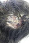
Due to the unfavourable response to treatment, she was referred to the dermatology department, where she received a diagnosis of tinea capitis and started topical treatment with a terbinafine cream and a sertaconazole nitrate gel. The relevant findings of the history were a past episode of tinea corporis in the father and the presence of a pet guinea pig in the household that also had skin lesions. Two weeks later, the lesions had progressed into abscesses (Fig. 1). The abscesses were actively drained every 48h, with collection of a sample of the exudate and dystrophic hairs for submission to the microbiology laboratory. The patient received a prescription for a new course of treatment with oral itraconazole (62.5mg/day) and a miconazole/hydrocortisone cream for 3 weeks.
Girl aged 8 years referred to the dermatology department for assessment of a bald plaque in the scalp more than 1 month after onset of symptoms (lesions initially developed in the left breast and spread to the scalp over time). During the history taking, she reported that her pet guinea pigs had developed bald patches before she contracted the disease. A sample was collected for culture and the patient received treatment with terbinafine at a dose of 125mg/day for 1 month combined with topical administration of a terbinafine cream in the morning and a mometasone furoate and acetylsalicylic acid cream at night.
The microbiology laboratory processed the samples of both patients (Fig. 2). In both cases, 15 days after submission the laboratory report indicated the presence of a fungus initially identified as a Trichophyton species. The definitive identification as Arthroderma benhamiae was achieved by the analysis of the sequence of the internal transcribed spacer (ITS) region of ribosomal RNA.
Arthroderma benhamiae usually causes mild infections that respond to topical treatment with ciclopirox, imidazole or terbinafine. However, cases with more extensive involvement and tinea capitis require treatment with oral antifungals. In patients with kerion celsi, early diagnosis and prompt initiation of treatment are of the essence due to the risk of scarring hair loss. There are few studies on the use of different antifungals to treat infections by this fungus. Most authors report use of terbinafine, griseofulvin, itraconazole or fluconazole for a minimum of 4–6 weeks,4 with favourable outcomes. Epidemiological data, such as the presence of pets, especially guinea pigs, are important clues for suspecting and correctly diagnosing this fungal skin infection.
Please cite this article as: Martín-Peñaranda T, Lera Imbuluzqueta JM, Alkorta Gurrutxaga M. Arthroderma benhamiae en pacientes con cobayas. An Pediatr (Barc). 2019;90:51–52.


