


We present the case of a male infant aged 16 days with an unremarkable maternal obstetric history, born at 38 + 6 weeks of gestation. He was admitted on account of 2 febrile temperature peaks of 38 °C and pain on mobilization of the right shoulder. After an ultrasound examination detected arthritis in the right shoulder, culture of surgical specimens confirmed the presence of Staphylococcus aureus.
Given the worsening symptoms and with the aim of assessing for the potential presence of additional foci of infection, a whole-body positron-emission tomography (PET)/computed tomography (CT) scan was performed (Biograph™ mCT 20 scanner, Siemens Medical Systems; Knoxville, TN, USA), including the extremities (Fig. 1), with an administered activity of 36.8 MBq/0.99 mCi of 18F-fluorodeoxyglucose (18F-FDG) (paediatric dose recommended by the European Association of Nuclear Medicine)1 and low-dose CT-based attenuation correction (80 kVp, 5 mAs).

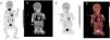
(A) Maximum intensity projection image. (B) Coronal CT image. (C) Coronal PET image. (D) Coronal PET/CT fusion image: 18F-FDG imaging with high uptake in the proximal epiphysis of the right humerus (blue arrow) associated with active osteomyelitis2 and moderate uptake in the articular surface of the right humeral head associated with the already established active articular illness.
Acute osteomyelitis, a rare complication in newborn infants, poses a diagnostic and therapeutic challenge.3 The value of 18F-FDG PET-CT in paediatric oncology is well established, although in children with fever or suspected infection, the evidence on the usefulness of 18FDG PET-CT imaging is scarce, despite its proven usefulness in the diagnosis of infectious or inflammatory foci in adult patients.
If there is uncertainty in the suspicion of infection and/or inflammation of synovial joints, the use of 18F-FDG PET-CT imaging could be contemplated in very select cases for the accurate assessment of septic arthritis with underlying acute osteomyelitis.
