


Naevoid acanthosis nigricans is considered a variant of epidermal naevus.1 It has also been referred to as “round and velvety epidermal naevus” (RAVEN) and “naevus resembling acanthosis nigricans.2
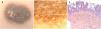
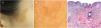
We present the cases of naevoid acanthosis nigricans in 2 adolescent patients aged 14 years (case 1) and 15 years (case 2), who sought care for an asymptomatic lesion at the dorsal level and right preauricular region, respectively, with onset 3 years prior. The examination revealed an oval hyperpigmented plaque with a velvety surface measuring 40 × 25 mm in case 1 (Fig. 1A) and 35 × 25 mm in case 2 (Fig. 2A). Dermoscopy revealed brown ovoid structures on a white background in case 1 (Fig. 1B) and a cerebriform appearance with fissures, crypts and round brown structures in case 2, (Fig. 2B). In both cases, the examination of the biopsy specimen evinced orthokeratotic hyperkeratosis, acanthosis, papillomatosis, hyperpigmentation of the basal layer and very mild superficial perivascular inflammatory infiltrate (Figs. 1C and 2C).
![(A) Oval, dark brown plaque with a velvety surface located in the back. (B) Dermoscopy revealed brown ovoid structures on a light background (FotoFinder medicam 800 HD digital epiluminescence system). (C) Orthokeratotic hyperkeratosis with acanthosis, papillomatosis and hyperpigmentation of the basal layer (haematoxylin-eosin [H-E] stain, original magnification ×40).](https://static.elsevier.es/multimedia/23412879/0000009900000005/v1_202311240521/S2341287923002326/v1_202311240521/en/main.assets/gr1.jpeg?xkr=ue/ImdikoIMrsJoerZ+w9/qVHBXBqbSQ7FNUvNof+6+l4v03CmyaR9Rm+q8TRfDERovN5wMUmDVyYvA+rI0XvOWA7j4/+5n1aGoKQ+OmCDYBzcuYrQFiPmIqiRFo8RWpl4O0fq3KGOssMk9qLtvsSNGxKuuViWNUMifaaAzu2Kz1YvExD6Qp9/P+CnOuViSy4wHMjbckRSuQlyy1pfb7P5M0Ob9zjXbAtcWvZiG4OGKE0AiKWiLVI3U0qe4klgpvGPNcvmllqdYsagWSy2x2RCwZI27LXeIxNE+lHfeGE9s=)
(A) Oval, dark brown plaque with a velvety surface located in the back. (B) Dermoscopy revealed brown ovoid structures on a light background (FotoFinder medicam 800 HD digital epiluminescence system). (C) Orthokeratotic hyperkeratosis with acanthosis, papillomatosis and hyperpigmentation of the basal layer (haematoxylin-eosin [H-E] stain, original magnification ×40).
(A) Oval, light brown plaque with a velvety surface located in the right preauricular region. (B) Dermoscopy allowed visualization of a cerebriform appearance with fissures and crypts (FotoFinder medicam 800 HD digital epiluminescence system). (C) The histological findings were the same as in case 1 (H-E stain, original magnification ×40).
Naevoid acanthosis nigricans is a rare condition, with fewer than 50 cases reported to date. The presence of mosaic mutations in genes FGFR2 and FGFR3 has been described in association with this disease.1 It manifests with hyperpigmented plaques with a velvety surface. It develops in childhood or adolescence, and the most frequent sites of the lesions are the trunk, head and extremities.1,3 Histologically, it resembles classic acanthosis nigricans. The differential diagnosis includes melanocytic naevus, epidermal naevus and linear verrucous epidermal naevus, among others.2 Topical vitamin D analogues and retinoids and local destructive procedures, such as laser ablation, are some treatment options, although conservative treatment is an equally appropriate alternative.2,3
FundingNone of the authors received any funding to carry out this project.
Previous meeting: this report was presented as a poster at the 34th Meeting of the Spanish Group of Paediatric Dermatology, January 27–28, 2023.
