Información de la revista
Vol. 98. Núm. 5.
Páginas 399-400 (Mayo 2023)
Images in Paediatrics
Acceso a texto completo
Brown spots on the tongue
Lesiones marrones en la lengua
Visitas
22374
Diego Soto-Garcíaa,b,
Autor para correspondencia
diego.soto.garcia@gmail.com

Corresponding author.
, Carmen Couselo-Rodrígueza,b, Sandra Martínez-Fernándeza,b, Ángeles Flóreza,b
a Servicio de Dermatología, Complejo Hospitalario Universitario de Pontevedra, Pontevedra, Spain
b Grupo de Investigación DIPO, Instituto de Investigación Sanitaria Galicia Sur (IIS Galicia Sur), SERGAS-UVIGO, Vigo, Spain
Este artículo ha recibido
Información del artículo
Texto completo
Bibliografía
Descargar PDF
Estadísticas
Figuras (2)
Texto completo

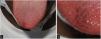
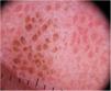
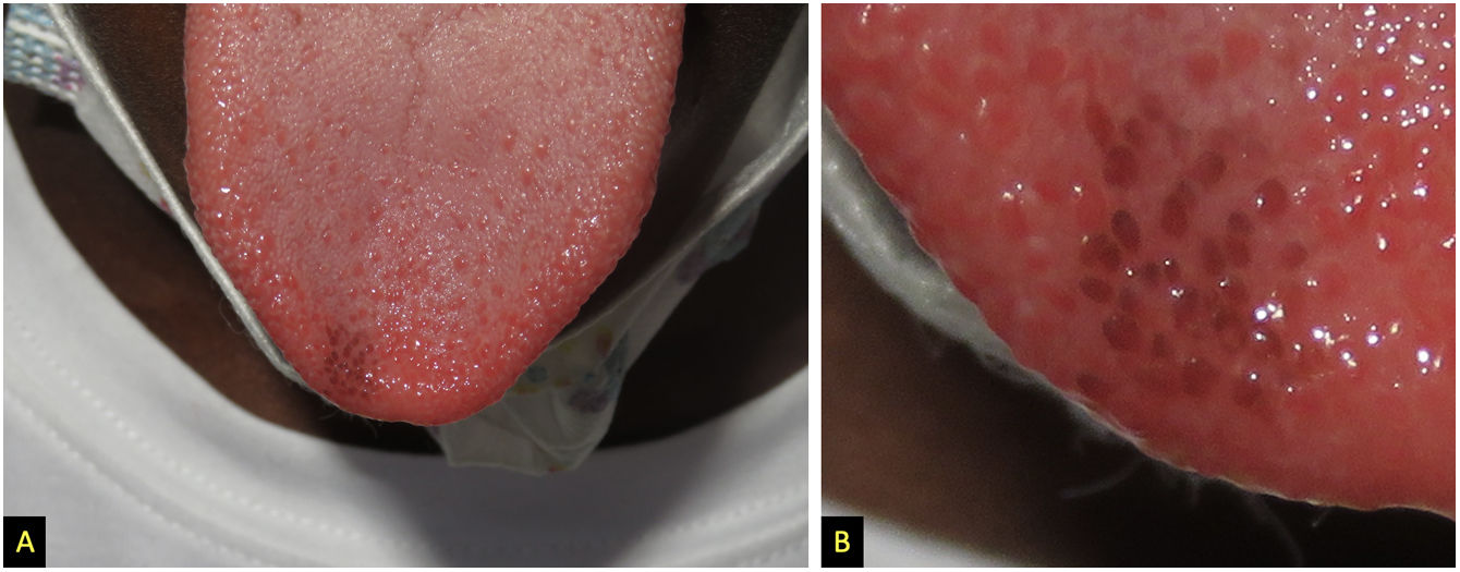
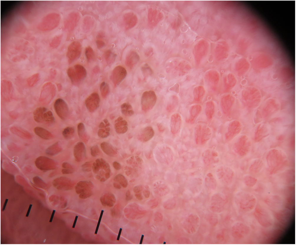
A boy aged 11 years of African ancestry presented with macules in the tongue with onset 2 years prior. The patient was asymptomatic and had not experienced any trauma, was not taking any medication and had no family history of cancer. The physical examination revealed multiple brownish macules measuring 1 mm in diameter in the papillae of the tip of the tongue (Fig. 1). Hyperpigmentation was not detected elsewhere. The examination with dermoscopy evinced multiple brownish projections dividing the vessels at the base (Fig. 2). Given the location, characteristic dermoscopic findings and lack of systemic manifestations, a diagnosis of pigmented fungiform papillae of the tongue was made without need of a biopsy.1,2

Figure 1.

A. Multiples brown fungiform papules located in the tip of the tongue. B. Detail.

Figure 2.

Dermoscopy (DermLite DL200; polarised light, 10× magnification). Multiple projections with pigmented borders with a “rose petal” appearance.

Pigmented fungiform papillae of the tongue is a benign and little-known condition that is more frequent in individuals of African descent and with high phototypes.1,3 The lesions are typically located in the tip or sides of the tongue. Hyperpigmentation is exclusively found in the papillae and usually has onset in childhood and is not progressive.2 The differential diagnosis includes Addison disease, Peutz-Jeghers syndrome, black hairy tongue, melanotic macule and melanocytic naevus.1 Paediatricians and paediatric dermatologists must be aware of this condition, as it can be diagnosed based on the clinical picture, avoiding the use of invasive tests.

References
[1]
C.L. Ross, C. Ring, S. Yang.
Pigmented fungiform papillae of the tongue.
JAMA Dermatol, 156 (2020), pp. 1249
[2]
M.A. Chessa, A. Patrizi, A. Sechi, A. Virdi, M. Leuzzi, I. Neri.
Pigmented fungiform lingual papillae: dermoscopic and clinical features.
J Eur Acad Dermatol Venereol, 32 (2018), pp. 935-939
[3]
S.M. Rice, K. Lal.
Hyperpigmented macules of the tongue in a 15-year-old girl.
Pediatr Dermatol, 38 (2021), pp. 941-942
Copyright © 2022. Asociación Española de Pediatría
Descargar PDF
Idiomas
Anales de Pediatría
Opciones de artículo
Herramientas