

The enlargement of the hilar periportal space manifests with what is known as a “periportal halo”, “periportal cuffing” or “periportal collar sign” on sonography.1 This is a very rare sign, with an incidence of 0.95%, which can be echo-rich (91% of cases) or echo-poor (9%) in appearance.1
Most cases of echo-poor periportal cuffing are associated with malignancy, especially with blood tumours, and are caused by transient lymphoedema resulting from the blockage of small lymph vessels or from direct periportal infiltration by malignant cells.2 The sonographic abnormality tends to disappear within 2 weeks of initiating chemotherapy.2,3
We present the case of a boy aged 18 months brought in by his parents for assessment of gastrointestinal symptoms, irritability and decreased appetite with onset 3–4 weeks prior.
The patient had been discharged from hospital 13 days before with a diagnosis of mesenteric adenitis.
The history-taking and physical examination revealed nonspecific malaise, pallor of the skin and mucosae, mild abdominal distension and haematomas in the groin and anterior surface of both lower extremities.
An abdominal ultrasound scan revealed hepatomegaly with periportal hypoechogenicity around the main portal vein and its branches (Figs. 1 and 2), and mild splenomegaly with multiple accessory spleens in the hilum (Fig. 3). The patient was referred to the corresponding referral hospital due to suspicion of myeloproliferative disease, where he received a diagnosis of acute myeloid leukaemia.
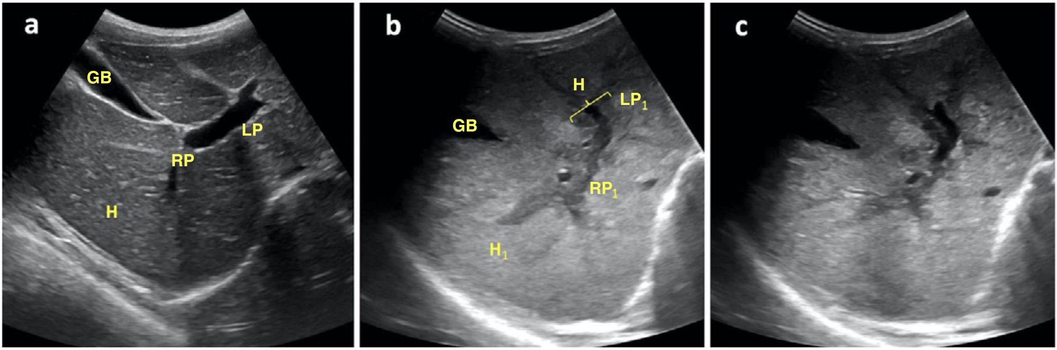
 Homogeneous echogenicity of hepatic parenchyma (H) with normal appearance of right portal vein (PD), left portal vein (PI) and gallbladder (VB) in a healthy patient; b) Heterogeneous echogenicity of hepatic parenchyma (H1), right portal vein (PD1) and left portal vein (PI1) with a hypoechoic halo (M) in patient with acute myeloid leukaemia; c) Similar image in the same patient, without references to allow better visualization of the aforementioned structures.")
Oblique transverse plane of the hypochondrium obtained with a convex probe at the level of the epigastrium, showing: a) Homogeneous echogenicity of hepatic parenchyma (H) with normal appearance of right portal vein (PD), left portal vein (PI) and gallbladder (VB) in a healthy patient; b) Heterogeneous echogenicity of hepatic parenchyma (H1), right portal vein (PD1) and left portal vein (PI1) with a hypoechoic halo (M) in patient with acute myeloid leukaemia; c) Similar image in the same patient, without references to allow better visualization of the aforementioned structures.
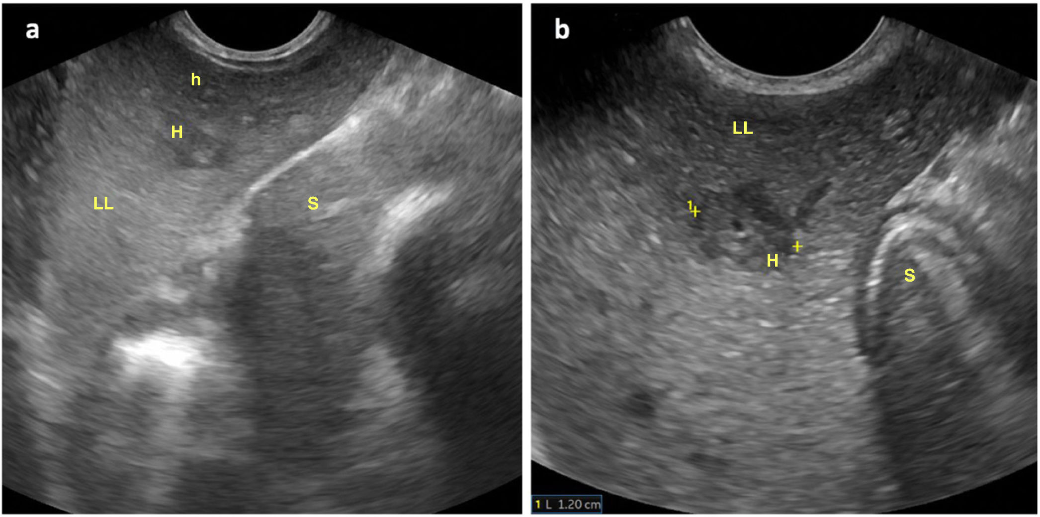
 Hepatic parenchyma at the level of the left lobe (LHI) with transversal sections of echo-poor periportal haloes of larger (M) and smaller (m) size; b) Enlargement of previous image with measurement of the diameter of one of the periportal haloes. Stomach (E).")
Oblique longitudinal plane of the left hypochondrium obtained with a convex probe at the level of the epigastrium, showing: a) Hepatic parenchyma at the level of the left lobe (LHI) with transversal sections of echo-poor periportal haloes of larger (M) and smaller (m) size; b) Enlargement of previous image with measurement of the diameter of one of the periportal haloes. Stomach (E).
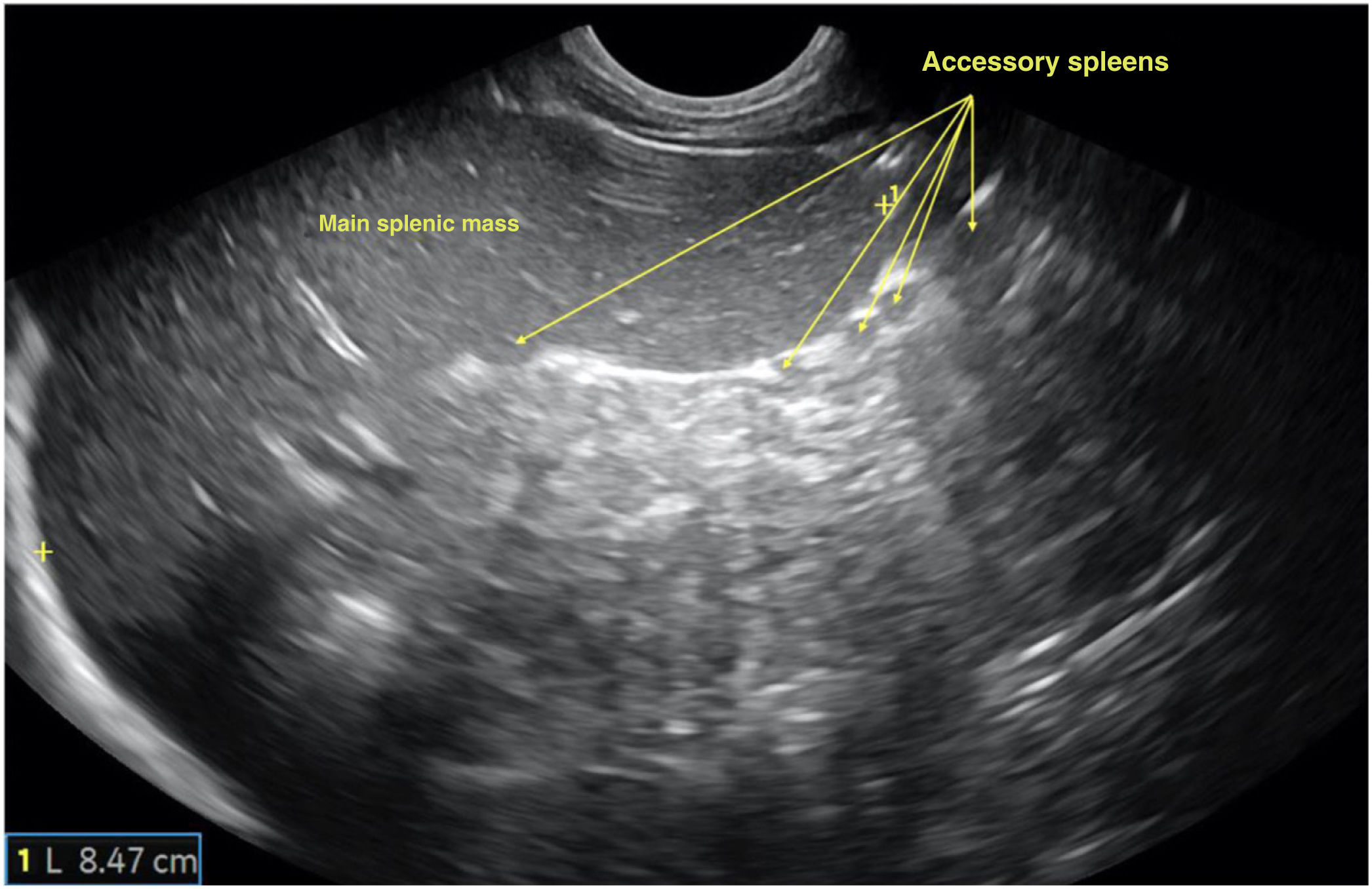
 and several accessory spleens in the lower pole of the hilum, suggestive of malignancy.")
Longitudinal plane of the spleen obtained with a convex probe at the level of the left hypochondrium evincing splenomegaly (length of long axis of spleen, 8.47cm; upper limit of normal for age, 8cm) and several accessory spleens in the lower pole of the hilum, suggestive of malignancy.
The compatible presentation and the identification of sonographic signs suggestive of a blood tumour allowed earlier diagnosis and treatment, thus contributing to the favourable outcome in this patient.

