


11β-hydroxylase (11β-OH) deficiency is the second most frequent cause of classic congenital adrenal hyperplasia (CAH) (5%–8% of cases). Clinically, it is characterized by virilization and arterial hypertension. The objective of this study was to describe the clinical, biochemical and genetic characteristics classic 11β-OH deficiency in patients managed in our hospital and its outcomes.
Patients and methodsRetrospective longitudinal, observational and descriptive study. Inclusion criteria: Patients with clinical features of virilization, high levels of 11-deoxycortisol and study of CYP11B1 gene with detection of pathogenic and likely pathogenic variants.
ResultsWe identified 6 patients (1 male, 5 female) from 4 families. In the 4 index cases, the median age at diagnosis was 2.3 years. The 46,XX patients exhibited a variable degree of virilization at diagnosis, with a predominance of Prader stage V, and one case of male sex assignment at birth. All patients had elevated serum concentrations of 17-hydroxyprogesterone and testosterone. Fifty percent of the patients had developed arterial hypertension during the follow-up, with onset at a median age of 9.3 years. Three 46,XX patients reached a median final height of 154 cm. Six different variants of theCYP11B1 gene were identified, 5 of which were novel variants (c.595 G > A, c.710 T > C, c.1156delG, c.395 + 2dupT, c.1159dupA).
ConclusionsThere is considerable heterogeneity in the clinical presentation of patients with CAH due to 11β-OH deficiency. Early diagnosis and treatment are important to prevent complications and improve long-term outcomes. We report 6 different variants of the CYP11B1 gene, including 5 novel variants.
La deficiencia de 11β-hidroxilasa (11β-OH) es la segunda causa más común de las formas clásicas de hiperplasia suprarrenal congénita (HSC) (5–8% de los casos). Clínicamente se caracteriza por virilización e hipertensión arterial. El objetivo de este estudio es describir las características clínicas, bioquímicas, genéticas y la evolución de los pacientes seguidos en nuestro centro.
Pacientes y métodosEstudio observacional longitudinal, retrospectivo y descriptivo. Criterios de inclusión: clínica de virilización, 11-desoxicortisol elevado en plasma y estudio del gen CYP11B1 con variantes patogénicas y probablemente patogénicas.
ResultadosSe identificaron 6 pacientes (1H, 5 M) pertenecientes a 4 familias. La edad media al diagnóstico de los 4 pacientes índice fue de 2.3 años. Las pacientes 46,XX mostraron grados variables de virilización al diagnóstico, predominando el estadio 5 de Prader, con un caso de asignación de género masculino al nacimiento. Todos los pacientes presentaron aumento de las concentraciones séricas de17-hidroxiprogesterona y testosterona. El 50% de los pacientes han desarrollado hipertensión arterial, con una mediana de edad de 9,3 años. Tres pacientes 46,XX han alcanzado talla final con una mediana de 154 cm. Se encontraron 6 variantes diferentes distribuidas a lo largo del gen CYP11B1, 5 de las cuales son variantes no descritas previamente (c.595 G > A, c.710 T > C, c.1156delG, c.395 + 2dupT, c.1159dupA).
ConclusionesLos pacientes con formas clásicas de HSC por déficit de 11β-OH presentan una gran heterogeneidad en la expresión clínica. El diagnóstico y el tratamiento precoces son importantes para prevenir complicaciones y mejorar los resultados a largo plazo.

Congenital adrenal hyperplasia (CAH) is an autosomal recessive disorder caused by defects in different enzymes involved in adrenal steroid hormone synthesis, most frequently 21-hydroxylase deficiency (21OHD), which accounts for 90%–95% of cases.1 The second most frequent cause is 11β-hydroxylase (11β-OH) deficiency (5%–8% of cases), with an estimated annual incidence of 1 per 100 000–200 000 live births in in nonconsanguineous populations that can increase to up to 1 per 5000 births in the population of Moroccan Jewish descent.2–5
11β-hydroxylase deficiency is caused by genetic changes in the CYP11B1 gene, located in chromosome 8q24.3. It exhibits a high sequence homology with the CYP11B2 gene (aldosterone synthase), which is approximately 40 kb apart.6,7 The CYP11B1 gene encodes an enzyme in the cytochrome P450 system responsible for the conversion of 11-deoxycortisol to cortisol and 11-deoxycorticosterone (DOC) to corticosterone.8,9 Approximately 180 pathogenic variants have been described to date; most of them are missense variants (60%) and they tend to cluster in exons 2, 6, 7 and 8.10–12
Phenotypically, there are two types of 11β-hydroxylase deficiency, the classic and nonclassic forms, differentiated by the degree of clinical severity and of enzymatic activity loss. The classic, more severe form is characterized by serious deficiency, with a residual enzymatic activity of less than 10% that results in reduced cortisol synthesis and a corresponding elevation in adrenocorticotropic hormone (ACTH), accumulation of glucocorticoid and mineralocorticoid precursors proximal to the enzymatic block and excessive androgen biosynthesis.3,13 Hyperandrogenism causes a varying degree of virilization of the external genitalia in affected girls and macrogenitosomia in affected boys, and patients of any sex can experience peripheral precocious puberty, premature epiphyseal closure and accelerated somatic growth. The accumulation of DOC and its metabolites, which have mineralocorticoid activity, causes sodium retention and volume expansion that in turn result in low-renin hypertension in approximately two thirds of patients.14,15
Compared to clinical studies of 21OHD, few studies have analyzed the clinical and hormonal phenotype 11β-OH deficiency, and few case series have been published due to the low frequency of the disease.
The aim of our study was to describe the clinical, biochemical and genetic characteristics and the outcomes of patients with classic 11β-OH deficiency managed in our hospital.
Patients and methodsWe conducted a retrospective observational study in a cohort of patients with a diagnosis of classic 11β-OH deficiency managed at the pediatric endocrinology unit of a tertiary care hospital between June 1996 and January 2022.
Inclusion criteria: clinical features of virilization, elevation of 11-deoxycortisol (>7.2 ng/mL) and sequencing of gene CYP11B1 in patients with likely pathogenic or pathogenic variants. Exclusion criteria: lack of follow-up data.
Retrieval of data recorded at diagnosis and during follow-up: chronological age (years), sex assigned at birth, relevant family history, consanguinity, clinical manifestations, findings of physical examination, laboratory test results, imaging, hydrocortisone dose (mg/m2/day), antihypertensive medication and associated complications. We expressed anthropometric data as z scores, calculated with reference data obtained in the Spanish population.16 The degree of genital virilization in 46XX patients was assessed with the Prader scale.17 Penile length (PL) was expressed in cm and percentiles.18 Pubertal stage was assessed according to the Marshall and Tanner staging system.19,20 We assessed bone age with the Greulich and Pyle method and calculated the mid-parental height (MPH) with the method proposed by Tanner.21 We defined hypertension (HTN) as an arterial blood level persistently above the 95th percentile for age, sex and height measured in at least 3 separate visits.22 Ambulatory blood pressure monitoring (ABPM) was performed with the Custo Screen Pediatric recorder in patients aged less than 12 years and the Custo Screen 310 recorded in patients aged more than 12 years and analyzed with the Custo Diagnostic software. The analysis was carried out in adherence with current guidelines for the interpretation of ABMS in the children and adolescents.23
The descriptive statistical analysis was carried out with the software SPSS, version 21.0. We summarized values as median and interquartile range (IQR).
ResultsWe identified six patients (1 male, 5 female) from four different families: family 1, of Romani ethnicity and with consanguineous parents, family 2, of Moroccan origin with nonconsanguineous parents, and families 3 and 4, with Caucasian nonconsanguineous parents.
None of the families had a known history of neonatal death or relevant endocrine disease.
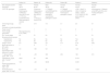
Clinical and biochemical characteristics at diagnosisTable 1 presents the clinical characteristics of the patients at the time of diagnosis. In the four index cases, the median age at diagnosis was 2.3 years (IQR 1.14–6.45). The other 2 cases, corresponding to younger siblings in families 1 and 2, were diagnosed at age 7 months (in a patient whose parents refused prenatal diagnosis) and prenatally, respectively. None of these 2 patients received prenatal treatment with dexamethasone, according to the wishes of the parents.
Clinical characteristics of patients with 11β-OH deficiency at the time of diagnosis.
| Patient 1A | Patient 1B | Patient 2A | Patient 2B | Patient 3 | Patient 4 | |
|---|---|---|---|---|---|---|
| Sex assigned at birth | Male | Female | Male | Female | Female | Female |
| Karyotype | 46,XY | 46,XX | 46,XX | 46,XX | 46,XX | 46,XX |
| CYP11B1 genotype | Compound heterozygous c.595G>A p.(Ala199Thr) and c.710T>C p.(Leu237Pro)Two likely pathogenic variants | Compound heterozygous c.595G>A p.(Ala199Thr) and c.710T>C p.(Leu237Pro) | c.1156delG (p.Ala386Argfs*44) homozygous pathogenic variantLikely pathogenic variant | c.1156delG (p.Ala386Argfs*44) homozygous pathogenic variant | c.395 + 2dupT in intron 2 (maternal allele) + hybrid CYP11B2/CYP11B1 gene (paternal allele) | Homozygousc.1159dupA; p.(Ser387Lysfs*35), exon 7Likely pathogenic variant |
| Chronological age, years | 2.4 | 0.74 | 7.8 | 0.1 | 2.4 | 0.1 |
| Virilization of external genitalia, | ||||||
| Prader scale(1) | – | 1 | 5 | 5 | 3 | 5 |
| Penile length (percentile) | Macrogenitosomia 6 cm (>95th) | – | – | – | – | – |
| PH, Tanner stage | P2 | P1 | P4 | P1 | P1 | P1 |
| Mineralocorticoid excess | ||||||
| Hypertension | No | No | No | No | Yes | No |
| Hypokalemia | No | No | No | No | No | No |
| Height z score | 5.4 | 0.99 | 3.5 | 0.11 | 3.78 | 0.92 |
| Bone age based on GP, years | 7 | 2 | 17 | – | 3.6 | – |
| Bone age-chronological age, difference in years | 4.6 | 1.26 | 9.2 | – | 1.26 | – |
| 11-deoxycortisol, ng/mL (NR, <7.2)a | 33 | > 59 | 333 | 254 | 128 | 213 |
| Testosterone, ng/dL (NR, 7−25.5)b | 49.61 | 53 | 429 | 214.9 | ||
| 17 Hydroxyprogesterone, ng/mL (NR, 0.05−1.6)b | 12.3 | > 20 | 92.86 | 43.08 | ||
| Androstenedione ng/mL (NR, 0.5−4.8)b | 8.2 | > 10 | 22 | 10.09 | ||
Abbreviations: GP, Greulich and Pyle method; NR, normal range; PH, pubic hair; SD, standard deviation.
Prader scale (1954): 1, clitoromegaly without labial fusion; 2, clitoromegaly and posterior labial fusion; 3, greater degree of clitoromegaly, single perineal urogenital orifice, and almost complete labial fusion; 4, increasingly phallic clitoris, urethra-like urogenital sinus at base of clitoris, and complete labial fusion; 5, appearance of male genitalia.
The 46,XY patient exhibited macrogenitosomia and premature pubarche. The five 46,XX patients exhibited varying degrees of virilization at diagnosis, three of them were classified as Prader stage 5 and one was assigned male sex at birth. In the latter, after diagnosis of CAH at age 7.8 years, the parents refused sex reassignment.
Advanced bone age was present at the time of diagnosis, with a median difference between bone age and chronological age of 2.9 years (IQR, 1.2–8.05). All patients had elevated serum levels of 11-deoxycortisol, 17-hydroxyprogesterone (17OHP) and testosterone. Only one patient had HTN at the time of diagnosis.
Genetic findingsIn all index cases, the CYP21A2 gene was sequenced first, and no pathogenic variants were found.
In the analysis of the CYP11B1gene (NM_000497.4), novel pathogenic variants were identified in three of the families.
In family 1, the analysis identified the changes c.595G>A (p.Ala199Thr) (affecting the last coding nucleotide of exon 3, therefore probably affecting splicing; ESEfinder and other tools [PolyPhen, SIFT] considered it pathological) and c.710T>C (p.Leu237Pro), considered pathological by different tools (PolyPhen, SIFT, MutationTaster). In family 2, analysis detected the homozygous variant c.1156delG (p.Ala386Argfs*44), also not previously described, a frameshift deletion predicted to result in a truncated protein and, therefore, loss of function. In patient 3, two compound heterozygous variants were identified: c.395 + 2dupT and a hybrid CYP11B2/CYP11B1 gene. The c.395 + 2dupT variant had not been previously described. In patient 4, there was a homozygous variant in exon 7, c.1159dupA (p.Ser387Lysfs*35), which also resulted in a reading frame shift.
All parents underwent genetic testing and were found to be carriers.
Medical treatmentAll patients were treated with hydrocortisone upon receiving the diagnosis of CAH, at a median dose of 12.6 mg/m2/day (IQR, 12.02–14.4). Over the course of treatment, the dose ranged between 10 and 20 mg/m2/day, except in patient 2A, who required higher doses of up to 27 mg/m2/day to normalize blood pressure.
Three patients (50%) had received fludrocortisone prior to diagnosis of CAH due to 11β-OH deficiency.
SurgeryThree 46 XX patients underwent genital reconstruction surgery: two at Prader stage 5 of virilization and the other at stage 3. The median age at the time of the first genital surgery was 10 months (5–37). Patient 2A underwent hysterectomy, bilateral salpingo-oophorectomy, resection of the upper third of the vagina and placement of a testicular prosthesis at age 8 years.
OutcomesThree patients have reached their adult height.
Patient 2A, with a 46,XX karyotype, male gender and late diagnosis at age 7.8 years reached a final height of 146 cm (2.81 SDs below the MPH).
The other two patients who have reached their final heights were diagnosed in the neonatal period. The birth length was normal for gestational age in both, while the height z scores at age 3 years, at the onset of peak height velocity and on reaching the final height were −1.2, −0.82 and −1.28 in patient 2B and −1.04, −1.51 and −1.61 in patient 4, respectively. Based on the age at the onset of PHV, patient 2B exhibited early maturation (onset at age 9 years) and patient 4 very early maturation (onset at age 8 years). Both patients exhibited decreased growth velocity in the first year of life, absence of prepubertal catch-up growth and normal growth after puberty. The final height was 1.02 SDs below the MPH for patient 2B and 0.55 SDs below the MPH for patient 4.
As regards blood pressure, three patients have developed HTN during the follow-up, which was detected at a median age of 9.3 years (IQR, 2.4–9.3). Patient 2A developed hypertension 8 years after diagnosis of CAH, which was attributed to poor adherence and required treatment with an antihypertensive drug (amlodipine). The two other patients in whom HTN was detected in the office were assessed with ABPM; patient 2B received a diagnosis of ambulatory nocturnal HTN, which was managed with dietary measures and adjustments to hormone therapy. In patient 3, monitoring detected severe HTN, with no target organ damage, which prompted an increase in the hydrocortisone dose that achieved marked improvement in blood pressure values. No other treatment has been initiated to date, and there has been no evidence of target organs have remained free of damage has been no evidence during the follow-up.
Last of all, the bone mineral density z score was normal in two patients (2A and 4).
Table 2 summarizes the clinical characteristics at the time of the last follow-up visit.
Clinical and biochemical characteristics of patients with 11β-OH deficiency at the time of the last follow-up visit.
| Patient 1A | Patient 1B | Patient 2A | Patient 2B | Patient 3 | Patient 4 | |
|---|---|---|---|---|---|---|
| Chronological age, years | 5.8 | 1.4 | 21 | 14.6 | 8.7 | 19 |
| Height (cm) | 124.6 | 80.6 | 146 | 155.2 | 128.6 | 156.5 |
| Height z score | 2.9 | 2.8 | −3.43 | −1.64 | −0.4 | −1.61 |
| Body mass index (kg/m2) | 18.1 | 18.3 | 25.7 | 29.8 | 16.7 | 21.9 |
| Diagnosis of HTN | No | No | Yes | Yes | Yes | No |
| Testosterone, ng/dL | <10 | 18.7 | NA | 11 | 12.6 | 34 |
| Androstenedione, ng/mL | 0.7 | 4 | <0.1 | 2 | 1.8 | 2.7 |
| Bone age, years | 13 | 2 | NA | NA | 11 | NA |
| Hydrocortisone dose (mg/m2/day) | 16 | 12.7 | 26.5 | 17 | 14.1 | 18.4 |
| Antihypertensive medication | No | No | Yes. amlodipine | No | No | No |
HTN, hypertension; NA, not applicable.
In this study, we describe the clinical, biochemical and molecular characteristics and long-term outcomes of 6 patients with classic CAH secondary to 11β-OH deficiency.
Since there is no routine newborn screening for it, the diagnosis of CAH due to 11β-OH deficiency is frequently delayed, especially in male patients, in whom it does not manifest with salt wasting.24 The median age at diagnosis of CAH in the four index cases was 2.3 years, similar to the median 2.8 years (range, 0–10) reported by Al-Jurayyan et al.25
As regards the clinical manifestations, the five 46,XX patients exhibited varying degrees of virilization at diagnosis, classified as Prader stage 5 in three. One of these patients, in whom the diagnosis of CAH was delayed, had been assigned male sex at birth. Some authors have reported that virilization tends to be more severe in female patients with 11β-OH deficiency compared to those with 21-OH deficiency and that it is more common for 46,XX newborns with 11β-OH deficiency to be mistakenly assigned male sex at birth.26,27 We ought to highlight that patient 1B in our study did not exhibit signs of virilization at birth but exhibited clitoromegaly by age 8 months.
Postnatally, hyperandrogenism manifests in both male and female individuals with rapid somatic growth and accelerated skeletal maturation, resulting in premature epiphyseal closure and short stature in adulthood. Although several studies have analyzed final height in patients with 21-OH deficiency, few have studied it in patients with 11β-OH deficiency. Previous studies have reported an abnormal final height in patients with 11β-OH deficiency, independently of the age at diagnosis and the clinical and biochemical control.3,15 During the follow-up, 3 of the 46,XX patients achieved their adult height, with a median of 154 cm (range, 146–154) and a median difference between the final height and the target height z scores of −1.02. Hochberg et al. reported that the final height of 11 female patients of Moroccan and Iranian ancestry with 11β-OH hydroxylase ranged between the 3rd and 5th percentiles, with a mean value of 153.1 cm (SD, 2.9 cm), 10 cm shorter compared to the general population.24 Baş et al. reported a final height 1.5 SDs below the mean in both male and female patients.10 Patient 2A, with karyotype 46,XX and a male phenotype and with a chronological age of 7.8 years at diagnosis achieved a final height of 146 cm (−2.81 SDs below the MPH). In a recent retrospective study that analyzed follow-up data for 46,XX patients with CAH who were raised male, 11 of the 38 patients who reached their final height had 11β-OH deficiency, and the median final height in this subset was 149.2 cm (range, 132.8–172).24
In our case series, two of the patients who reached their final height had been diagnosed in the neonatal period. When we analyzed their growth, we found that, similar to what happens in 21-OH deficiency, both of them had a decreased growth velocity in the first year of life, which may have been related to use of higher doses of hydrocortisone.28 They did not exhibit catchup growth in the prepubertal period, and had a normal pubertal growth spurt (no difference in the height z score at the onset of peak high velocity compared to the final height).
Hypertension is one of the characteristic manifestations of 11β-OH deficiency. Historically, mild to moderate HTN has been reported in two thirds of cases at the time of diagnosis13,15; however, recent studies have found that its frequency ranges between 53.6% and 59%.10,26 Hypertension usually develops during childhood or adolescence, although it can also develop in the early years of life.11 In our cohort, 3 of the patients developed HTN, in one, it was detected at diagnosis, and in the other two during the follow-up. In two of these patients, adjusting the dose of hydrocortisone sufficed to normalize blood pressure values, evincing that there is a degree of correlation between metabolic control and HTN. Patient 2A was the only one who required initiation of antihypertensive medication, which was attributed to poor adherence to treatment and other predisposing factors, such as obesity. It has been hypothesized that hypertension results from the excess of DOC and DOC metabolites with mineralocorticoid activity, which would result in sodium retention and volume expansion and thus cause low-renin hypertension; however, not all patients develop HTN and there also is no correlation between plasma DOC levels and the severity of HTN.26,29 Some authors have proposed that HTN may result from increased tissue sensitivity to DOC or the presence of other metabolites with more pronounced mineralocorticoid activity.25 Another aspect worth mentioning is the lack of a clear correlation between virilization and the features of mineralocorticoid excess. Some female patients with severe virilization have normal blood pressure values, while patients with mild virilization may develop severe HTN causing target organ damage.11,30 Our study also found no correlation between the degree of virilization and the presence of HTN. Al-Jurayyan et al. described the cases of four siblings that presented with hypertension and hypokalemia, relatively low levels of 11-deoxycortisol and mild virilization that persisted despite adequate treatment with hydrocortisone.25
To date, more than 180 pathogenic variants of the CYP11B1 have been reported, most of which give rise to the classic form of 11β-OH deficiency.10,30 Contrary to 21-OH deficiency, the genotype-phenotype correlation in 11β-OH deficiency has yet to be fully established, as molecular and genetic tests for 11β-OH are relatively scarce and in many cases the functional impact of identified pathological variants of CYP11B1 has not been studied. According to several reports, clinical severity and biochemical characteristics may differ significantly even among members of the same family with the same genotype, which is indicative of phenotypic variability.25,31 In the cohort under study, we found 6 different variants distributed along the CYP11B1 gene, 5 of which had not been described before (c.595G>A, c.710T>C, c.1156delG, c.395 + 2dupT, c.1159dupA). In patient 3, genetic testing identified two compound heterozygous variants: c.395 + 2dupT and the hybrid gene CYP11B2/CYP11B1. The c.395 + 2dupT variant was novel. According to the interpretation of the ESE-Finder in silico tool, this novel variant affects the splice donor site. Similar pathogenic changes have been reported before: in 2018, Baş et al. described the c.395 + 3A>G variant in a male patient.10 To date, there have been very few reported cases of CYP11B2/CYP11B1 hybrid genes.32–35 This phenomenon was first described in 2001 by Portrat et al., who identified a homozygous hybrid gene containing the promoter and exons 1–6 of CYP11B2 and exons 7–9 of CYP11B1.32 Functional studies would be needed to elucidate the pathogenicity of these variants.
In conclusion, 11β-OH is a rare cause of CAH. Patients with the classic form of disease exhibit substantial phenotypic heterogeneity with no clear genotype-phenotype correlation, contrary to 21-OH deficiency. Early diagnosis and treatment are important in order to prevent complications and improve long-term outcomes. We also reported six different CYP11B1 variants, five of which had not been previously described, in addition to their correlation to clinical and biochemical findings, expanding on the previously known spectrum of pathogenic CYP11B1 variants and contributing to the improvement of genetic counseling.
Declaration of Generative AI and AI-assisted technologies in the writing processNo form of artificial intelligence was used in the writing of this article.
FundingThis research did not receive any specific grant from funding agencies in the public, commercial, or not-for-profit sectors.
Previous meeting: This study was presented as an oral communication at the 44th Congress of the Sociedad Española de Endocrinología Pediátrica; May 11–13, 2022; Oviedo, Spain.
