
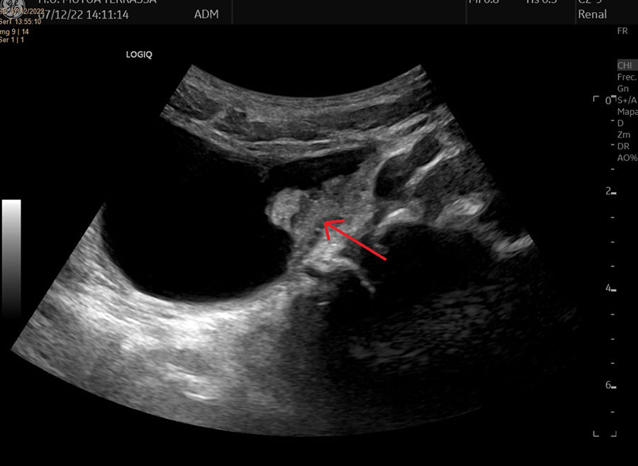
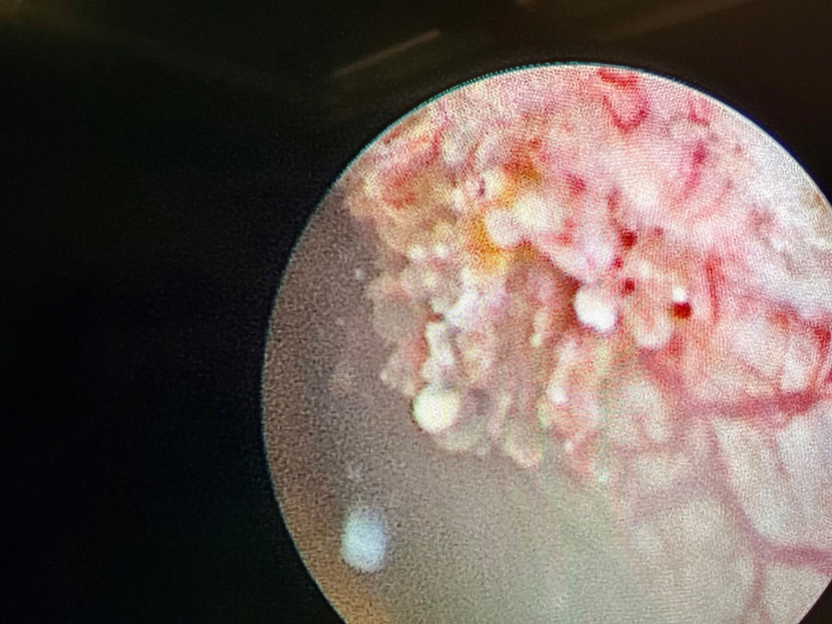
We present the case of a boy aged 6 years with chance finding of an intravesical mass in a follow-up ultrasound scan conducted 5 years after ureteral reimplantation surgery for vesicoureteral reflux. As seen in Fig. 1, the sonogram showed a hyperechoic parietal lesion measuring 10 × 5 mm and with well-demarcated borders, as well as evidence of vascularization in colour Doppler imaging. The scan also detected a second lesion of similar characteristics. A cystoscopy was performed, leading to detection and resection by diathermy of 4 lesions, including the 2 previously identified in the left inferolateral and posterior surfaces of the bladder and 2 smaller lesions located in the dome. Fig. 2 shows the largest one, located in the left inferolateral surface. The final histological diagnosis was nephrogenic adenoma of the bladder. Fig. 3 presents histological examination images including haematoxylin-eosin and immunohistochemistry staining. The patient remains asymptomatic and has not experienced recurrence one year after the intervention. On occasion, the routine follow-up of paediatric urologic patients gives rise to chance findings, and it is important to be aware of certain infrequent diseases that need to be included in the differential diagnosis. Nephrogenic adenoma is a metaplasia of the urothelium that may develop anywhere in the urinary tract.1 It is a benign condition, and it has been hypothesised that local inflammation or repetitive trauma could play a role in its development.2 It may be asymptomatic or manifest with dysuria, haematuria or recurrent urinary tract infection. The standard of care is complete endoscopic excision, and follow-up of the patient is required to detect potential recurrences.3


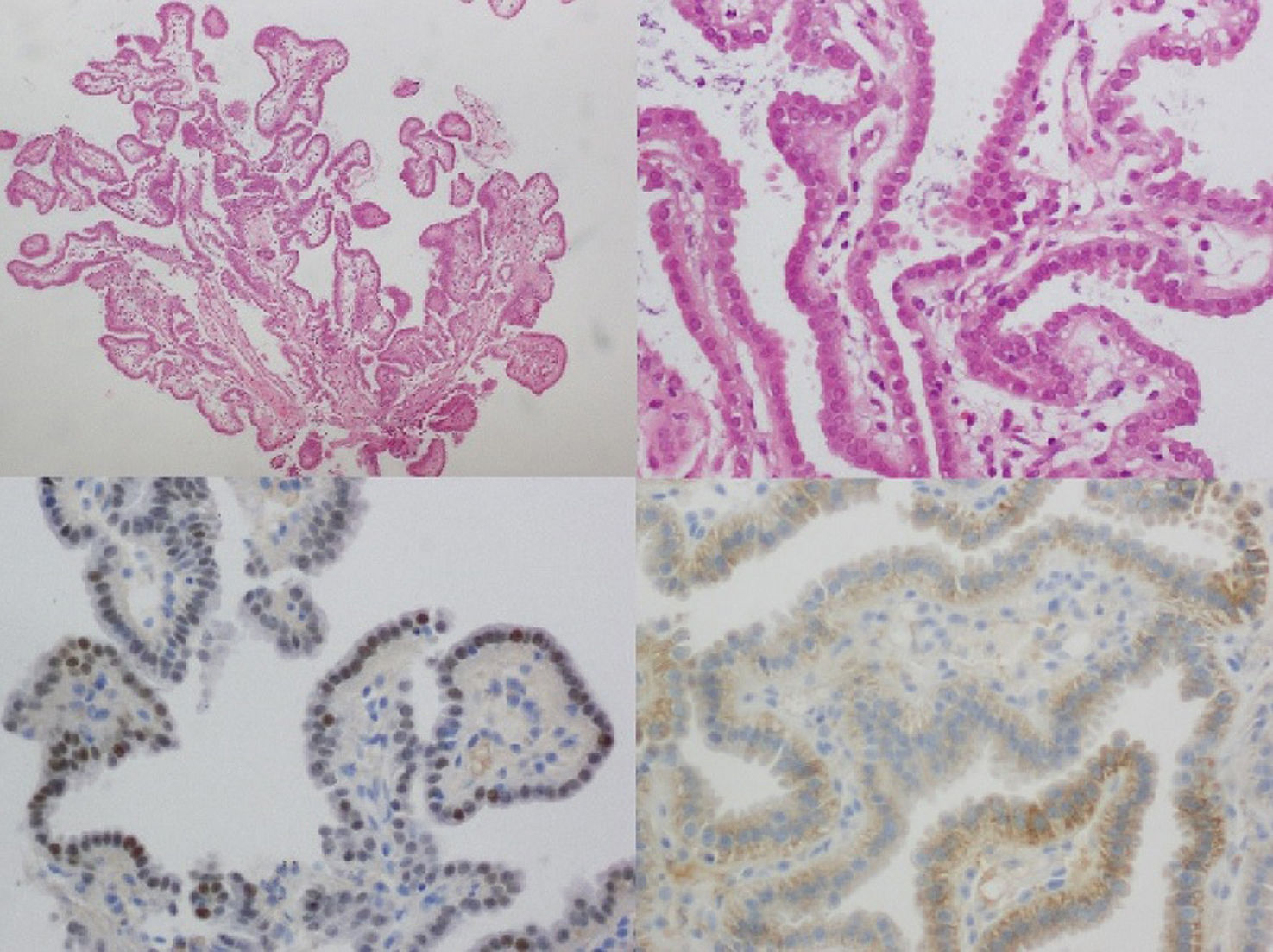
. Positive staining for α-methylacyl-CoA-racemase (bottom left) and PAX8 (bottom right).")
Histological examination. Papillary fronds lined with cuboidal epithelium with eosinophilic cytoplasm and smudged chromatin nuclei (haematoxylin-eosin stain, ×20 magnification on top left and ×40 on top right). Positive staining for α-methylacyl-CoA-racemase (bottom left) and PAX8 (bottom right).