Información de la revista
Vol. 99. Núm. 3.
Páginas 219-220 (Septiembre 2023)
Images in Paediatrics
Acceso a texto completo
Limp, petechiae and gingival hypertrophy: Think of scurvy
Cojera, petequias e hipertrofia gingival: pensar en escorbuto
Visitas
3796
Lara González-Garcíaa,
Autor para correspondencia
laragloria.gonzalez@sespa.es

Corresponding author.
, Begoña Fernández Martínezb, Carmen González Méndezc, Javier Gonzalez-Garciaa
a Hospital Universitario de Cabueñes, Gijón, Asturias, Spain
b Hospital Vital Álvarez-Buylla, Mieres, Asturias, Spain
c Hospital del Oriente de Asturias, Arriondas, Asturias, Spain
Este artículo ha recibido
Información del artículo
Texto completo
Bibliografía
Descargar PDF
Estadísticas
Figuras (3)
Mostrar másMostrar menos
Texto completo

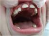
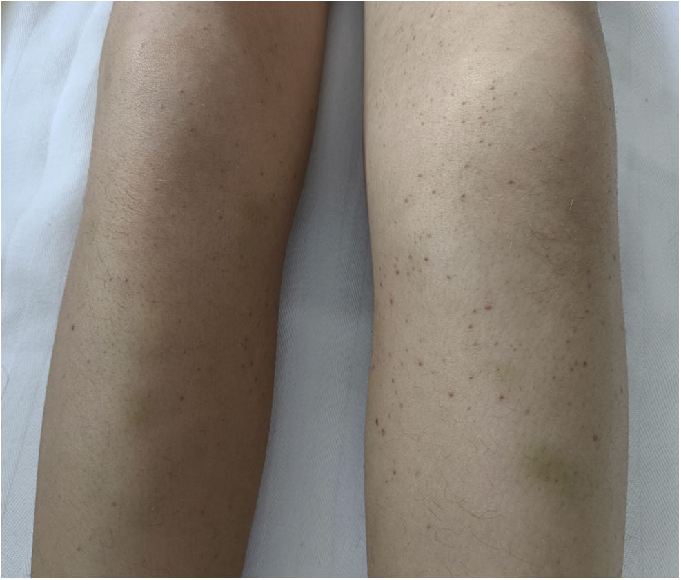
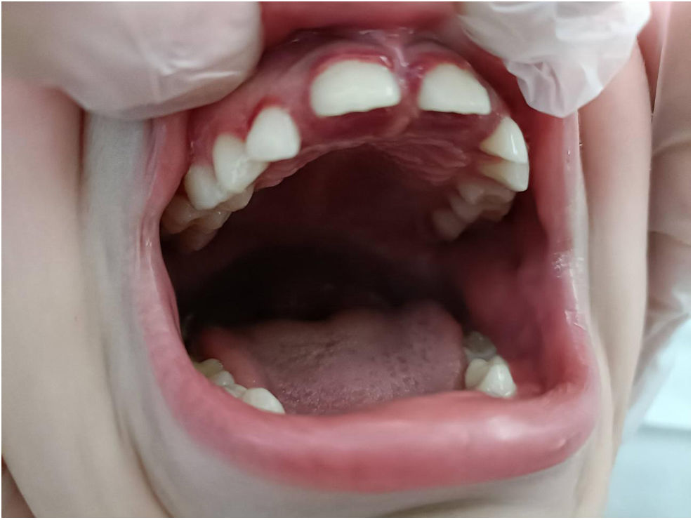
A boy aged 6 years presented with petechiae in the lower extremities, gingival enlargement with episodes of gum bleeding and inability to stand of 2 weeks’ duration. In addition, the patient had been unable to walk in the past 2 months due to bone pain in alternating lower extremities. The patient reported no pain on sitting, but did feel pain with passive mobilization of the lower extremities (Figs. 1–3).

Figure 1.

Plain radiograph of the left knee. Small bilateral metaphyseal spurs (green arrow), widening of the physeal lines, clear metaphyseal band (black arrow) in the distal femur and proximal tibia in the Trummerfel zone and white line of Frankel.

Figure 2.

Petechiae and perifollicular haemorrhages in the lower extremities associated with ecchymosis.

Figure 3.

Gingival hypertrophy.

The salient features of the personal history were diagnosis of autism spectrum disorder level 2, with hypersensitivity to textures and a very restrictive diet (lacking fruit and vegetables since age 3 years).

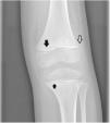
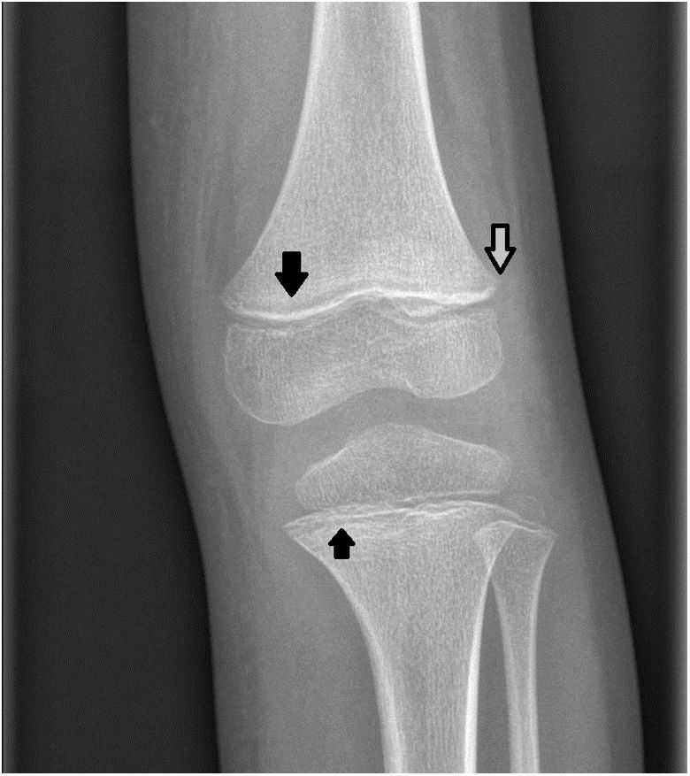
Laboratory tests revealed anaemia (haemoglobin concentration, 10.6 ng/dL) and an elevated erythrocyte sedimentation rate (61 mm/h), and normal results in the rheumatology screening tests. The hip and femur radiographs, ultrasound examination of the hip and the bone scintigraphy were normal, while the finding of the radiograph of the left knee were suggestive of scurvy.

The patient started treatment with ascorbic acid (300 mg/day for 7 days followed by 100 mg/day for 1 month) and improved rapidly, with full resolution of symptoms and normalization of test results within 2 weeks.

Scurvy must be considered in the differential diagnosis of patients presenting with petechiae, ecchymosis, gingivitis and joint pain after a few months of deficient vitamin C intake. It should also be suspected in patients with neurologic disorders who have restrictive diets.1–3

References
[1]
O. Iamopas, S. Ratanachu-Ek, S. Kaewnimee.
Scurvy in children — A neglected disease?.
Pediatr Int, 64 (2022),
[2]
P. Kothari, A. Tate, A. Adewumi, L.M. Kinlin, P. Ritwik.
The risk for scurvy in children with neurodevelopmental disorders.
Spec Care Dentist, 40 (2020), pp. 251-259
[3]
W.G. Sharp, R.C. Berry, L. Burrell, L. Scahill, B.O. McElhanon.
Scurvy as a sequela of avoidant-restrictive food intake disorder in autism: A systematic review.
J Dev Behav Pediatr, 41 (2020), pp. 397-405
Copyright © 2023. Asociación Española de Pediatría
Descargar PDF
Idiomas
Anales de Pediatría
Opciones de artículo
Herramientas