Sugerencias
Compartir
Información de la revista
Vol. 103. Núm. 4.
(Octubre 2025)
Images in Paediatrics
Acceso a texto completo
Giant borderline mucinous cystoadenoma of the ovary
Cistoadenoma mucinoso borderline gigante de ovario
Visitas
698
María Ruiz Medinaa,b,
Autor para correspondencia
mariaruizmedina.97@gmail.com

Corresponding author.
, Fernando Vázquez Ruedaa,b, María Rosa Ibarra Rodrígueza,b, Rosa Ortega Salasa,b, Rosa María Paredes Estebana,b
a Unidad de Gestión Clínica de Cirugía Pediátrica, Hospital Universitario Reina Sofía, Córdoba, Spain
b Unidad de Gestión Clínica Anatomía Patológica, Hospital Universitario Reina Sofía, Córdoba, Spain
Este artículo ha recibido
Información del artículo
Texto completo
Bibliografía
Descargar PDF
Estadísticas
Figuras (3)
Mostrar másMostrar menos
Texto completo

The incidence of ovarian tumors is low in the pediatric age group, accounting for around 1%–5% of solid tumors. In contrast to adults, the most common ovarian tumors in children are germ cell tumors, and cases of borderline cystadenoma are rare.1,2

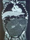
We present the case of a female patient aged 13 years with a giant borderline mucinous cystadenoma of the ovary. The patient presented with abdominal pain, nausea, vomiting and a palpable mass. The sonographic and CT findings suggested cystadenoma rather than cystadenocarcinoma (Fig. 1). Tumor markers were normal. The patient underwent a midline laparotomy and left salpingo-oophorectomy, which revealed a multilocular cystic neoplasm of irregular consistency and mucinous content weighing 7 kg (Figs. 2 and 3). The microscopic examination of the specimen revealed several solid foci of atypical cells compatible with borderline mucinous cystadenoma, as it met the criteria of nuclear stratification into two to three layers but without stromal invasion.3 We must underscore the importance of the histological analysis in the definitive diagnosis, as it allows differentiation between benign, borderline and malignant tumors. The cytologic evaluation was negative and the postoperative outcome was favorable, without need of adjuvant treatment.

Figure 1.

Coronal CT scan: multilocular trabeculated cystic mass measuring 34 cm × 25 cm × 16 cm.

Figure 2.

Intraoperative image: giant ovarian mass occupying the entire peritoneal cavity.

Figure 3.

Gross appearance: multilocular cystic mass with mucinous contents and solid foci revealed upon cutting.

Borderline mucinous cystadenoma is a rare tumor in children that clinicians ought to consider in the presence of an ovarian mass, as early diagnosis and correct surgical planning enable satisfactory outcomes.

References
[1]
F. Vázquez Rueda, F.J. Murcia Pascual, A. Siu Uribe, R.M. Ortega Salas, Á Escassi Gil, J.I. Garrido Pérez, et al.
Análisis de los tumores sólidos ováricos pediátricos en nuestra población.
An Pediatr., 92 (2020), pp. 88-93
[2]
F.E. Al Jama, A.A. al Ghamdi, T. Gasim, S.A. al Dakhiel, J. Rahman, M.S. Rahman.
Ovarian tumors in children and adolescents - a clinical study of 52 patients in a university hospital.
J Pediatr Adolesc Gynecol., 24 (2011), pp. 25-28
[3]
M. Iwasaki, K. Taira, H. Kobayashi, T. Saiga.
Ovarian mucinous cystadenoma of borderline malignancy in a premenarchal girl.
J Pediatr Adolesc Gynecol, 23 (2010), pp. e119-23
Descargar PDF
Idiomas
Anales de Pediatría
Opciones de artículo
Herramientas