


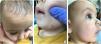

We present the case of a male infant aged 7 months that presented with well-demarcated, smooth yellow papules 3 to 5mm in size in his forehead, cheeks and scalp with onset 2 months prior (Fig. 1a–c). A skin lesion biopsy showed a small subepidermal proliferation of histiocytic cells extending into the papillary dermis. The cells were CD68 positive and CD1a and S100 negative (Fig. 2a–e).


a) Haematoxylin and eosin (H&E) staining (40×). The biopsy revealed a small subepidermal proliferation of cells extending into the papillary dermis and surrounded by an epidermal collarette. b) H&E staining (400×). The cells are histiocytic, with euchromatic nuclei, irregular contours and microvesicles in the cytoplasm. Slight fibrosis of the stroma, often in association with eosinophils. c) CD68 immunohistochemistry staining (200×). Cells positive for CD68. d) CD1a immunohistochemistry staining (200×). Cells negative for CD1a. e) S100 immunohistochemistry staining (200×). Cells negative for S100.
The patient received a diagnosis of benign cephalic histiocytosis (BCH). The immunohistochemistry findings were relevant for exclusion of Langerhans cell histiocytosis (LCH), the main differential diagnosis. Benign cephalic histiocytosis is a rare type of non-Langerhans cell histiocytosis characterized by the appearance of yellow to red-brown, asymptomatic macules and/or papules 2 to 5mm in size predominantly on the face, ears and neck.1 Skin lesions may develop between ages 3 and 36 months and resolve spontaneously over a period of months or years.
The pathogenesis is not yet fully understood. The differential diagnosis includes LCH, urticaria pigmentosa and the non-LCH disorders, especially juvenile xanthogranuloma (JXG) and generalized eruptive histiocytoma.
The most typical histologic pattern is a well-demarcated infiltrate of histiocytes in the superficial to mid-reticular dermis, expressing typical non-LCH histiocytic markers including CD11b/c, CD14b, CD68, HAM56, and factor XIIIa. Touton giant cells are more characteristic of JXG, although they may also be present in BCH.1,2 The number and distribution of lesions facilitates the diagnosis.
Since it is a self-limiting disorder contained to the skin, BHC does not require specific treatment.3
