


Preterm ovarian hyperstimulation syndrome is a rare condition characterized by swelling of the genitalia, clitoromegaly, elevated levels of follicle-stimulating hormone (FSH), luteinizing hormone (LH) and estradiol and follicular cysts.1,2 It is associated with the immaturity of the hypothalamic-pituitary-gonadal axis and diminished sensitivity of negative feedback from placental steroids, resulting in ovarian hyperstimulation.3 Given its very low incidence, with few described cases to date, reporting new cases is key to increase our knowledge of this condition and optimize its management.
We present the case of a girl born preterm at 23+5 weeks of gestation and appropriate for gestational age. At 62 days post birth (32+4 weeks of postmenstrual age), the girl presented with hard, swollen genitalia and clitoral enlargement (Fig. 1).
![Clinical course: substantial swelling in the hypogastric and genital areas with enlargement of the labia majora and clitoris, which improved during follow-up. (a) Age 3 months (36+3 weeks of postmenstrual age). (b) Age 4 months (2 weeks of corrected age [CA]). (c) Age 8 months (4 months of CA).](https://static.elsevier.es/multimedia/23412879/unassign/S2341287925004314/v1_202601120423/en/main.assets/gr1.jpeg?xkr=ue/ImdikoIMrsJoerZ+w9/qVHBXBqbSQ7FNUvNof+6+l4v03CmyaR9Rm+q8TRfDE6hnDWXuofgWkIZAAT/TS/ZCE46q/t7qhp1ZXZoKQ4GTrR8K7p1c8dEBIeH0fAAhWzjsA9sK43Y6jcheXJ17o/dPv9wNRQnoeQaELnx07BgmBMESvnu66SA29nmBNEahJgCT2RAgw7dQ03wokxIYTfJRLh8zOOTqnsPnJgJvEy182jHIsXPObWvY//38XIpzJ+fnbq8OK6QG7WqnjbDl1q9g/Ff8deAOU1t0GOk5UBr0n1yrrm2olHd6nkLQxsDXzcGkW61tX7GIsoX3bbweGNQ== "Clinical course: substantial swelling in the hypogastric and genital areas with enlargement of the labia majora and clitoris, which improved during follow-up. (a) Age 3 months (36+3 weeks of postmenstrual age). (b) Age 4 months (2 weeks of corrected age [CA]). (c) Age 8 months (4 months of CA).")
Clinical course: substantial swelling in the hypogastric and genital areas with enlargement of the labia majora and clitoris, which improved during follow-up. (a) Age 3 months (36+3 weeks of postmenstrual age). (b) Age 4 months (2 weeks of corrected age [CA]). (c) Age 8 months (4 months of CA).
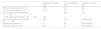
The blood work detected elevation of FSH, LH and estradiol levels (Fig. 2), and the abdominal ultrasound scan revealed ovarian enlargement and cystic lesions (Table 1).
 Left ovary (LO) at 3 months with a volume of 3.2cm3. (b) Right ovary (RO) at 3 months with a volume of 6.6cm3. (c) LO at 4 months with a volume of 2.2cm3. (d) RO at 4 months with a volume of 2.1cm3.")
Changes in laboratory and sonographic features.
| 3 months (36+3 weeks PMA) | 4 months (2 weeks CA) | 8 months (4 months CA) | |
|---|---|---|---|
| FSH (U/L) (normal range, 0.4−1.5) | 9.99 | 4.7 | 4.2 |
| LH (U/L) (normal range, 0.3−1.3) | 14.92 | 7.8 | 1.5 |
| 17β-estradiol (pg/mL) (normal range, 7.3±8.3) | – | 332 | 104 |
| 17-OHP (ng/mL) (normal range, 1.25±3.92) | 1.75 | – | – |
| Testosterone (ng/mL) (normal range, 0.68±0.08) | 0.68 | 0.58 | Undetectable |
| Left ovary ultrasound (cm3) | 3.2 | 2.2 | Not visualized |
| Right ovary ultrasound (cm3) | 6.6 | 2.1 | Not visualized |
Abbreviations: CA, corrected age; FSH, follicle-stimulating hormone; LH, luteinizing hormone; PMA, postmenstrual age; 17-OHP, 17-hydroxyprogesterone.
Elevation of FSH, LH and estradiol. Levels normalized at 11 months (7 months of CA). All other hormone levels measured in the patient were in the normal range.
Karyotyping was initially considered for the evaluation of ambiguous genitalia, but it was not performed on account of the clinical course and outcomes, which were consistent with preterm ovarian hyperstimulation syndrome.
The condition resolved spontaneously, with normalization of hormone levels and regression of cysts by age 11 months (7 months of corrected age).
In conclusion, preterm ovarian hyperstimulation syndrome is a rare, benign, and self-limiting condition. Although it usually does not require intervention, close clinical and ultrasound monitoring is recommended to prevent complications (cyst rupture or torsion) and avoid potential iatrogenic effects from unnecessary procedures.
FundingThis research did not receive any external funding.
