
The new coronavirus (SARS-CoV-2) that causes a severe acute respiratory syndrome emerges in Wuhan, China, in December 2019. It produces the aforementioned disease due to coronavirus 2019 (COVID-19), and has led to a declaration of a world public health emergency by the World Health Organisation. This new SARS-CoV-2 virus could share characteristics and an immune response similar to those described for other coronavirus. Given its activity on the interferon pathway, and the manner in which it dysregulates innate immunity, the use of treatments directed at modulating or containing this could be of interest. A narrative review was made of the current evidence about immunity against coronavirus and its applicability to SARS-CoV-2. The physiopathogenesis is also described, along with the underlying leucocyte activity, with the intention of clarifying the possible usefulness of inflammatory biomarkers and the development of personalised treatments.
El nuevo coronavirus (SARS-CoV-2) productor de síndrome respiratorio agudo severo surge en Wuhan, China, en diciembre de 2019. Genera la denominada enfermedad por coronavirus 2019 (COVID-19) y ha llevado a la declaración de emergencia de salud pública global a la Organización Mundial de la Salud. Este nuevo virus SARS-CoV-2 podría compartir características y respuesta inmune a las descritas para otros coronavirus. Dada su actividad sobre la vía del interferón y el modo en el que disregula la inmunidad innata, el uso de tratamientos dirigidos a modular o contener esta pueden ser de interés. Se realiza una revisión narrativa de la evidencia actual acerca de la inmunidad sobre coronavirus y su aplicabilidad para SARS-CoV-2. Se describe también la fisiopatogenia y la actividad leucocitaria subyacente, con intención de clarificar la posible utilidad de biomarcadores inflamatorios y el desarrollo de tratamientos personalizados.
The novel coronavirus, known as severe acute respiratory syndrome coronavirus 2 (SARS-CoV-2), emerged in Wuhan in December 2019. It causes the illness known as coronavirus disease 2019 (COVID-19) and has been declared a public health emergency of international concern by the World Health Organisation (WHO).1 At the time of this writing, there is a recognised global pandemic caused by this virus, with more than 185000 confirmed cases and 19500 deaths in Spain alone.2
Based on the data to date, COVID-19 is more prevalent and severe in elderly men with underlying conditions (high blood pressure, type 1 diabetes mellitus or cardiovascular disease).3–5 Attempts have been made to explain this fact from different perspectives. One of them is the immunological perspective, with the hypothesis that this may result from a faulty and insufficient response in individuals with a weakened immune system. Thus, responses that depend on innate or adaptive immunity could have a deleterious effect on the outcomes of disease.6,7
While the pathophysiology of COVID-19 has yet to be fully established, previous epidemics caused by other coronaviruses can help develop hypothesis about potential immune responses and thus anticipate them.6–8 Thus, both acute respiratory distress syndrome (SARS, caused by SARS-CoV) and Middle East respiratory syndrome (MERS, caused by MERS-CoV) were characterised by eliciting immune responses associated with the production of high levels of proinflammatory cytokines.6,9,10 Both diseases manifested with inflammation of the lungs, extensive lung damage and respiratory distress syndrome.11 They also presented with macrophage activation syndrome and immune dysregulation.6,7,12,13
In the present text, we provide a narrative review of the current literature on coronaviruses, SARS-CoV-2 and the immune system. We performed a structured search of articles indexed in PubMed. We searched for the following terms: “coronavirus”, “immunity” and “cytokines”. Due to the low number of hits, we also searched for the following combined terms: SARS-CoV-2, COVID-19 and “immunity”, “therapy” and “prognosis”. The search included articles published up to April 6, 2020. We then excluded all clinical guidelines or works whose abstracts did not refer to data relevant to our review. We included articles in English.
Coronavirus: characteristics and backgroundCoronaviruses are enveloped viruses with an unsegmented single-stranded RNA genome 26 to 32 kilobases in length. They belong to family Coronaviridae and order Nidovirales. They are widely distributed in humans and other mammals. Although in most cases infection by coronaviruses is mild, the SARS-CoV and MERS-CoV epidemics caused approximately 10000 deaths in the last 2 decades. The fatality rate of SARS has been estimated at 10% and the fatality rate of MERS at up to 37%.6,9,10
The SARS-CoV-2 generally causes mild or cold-like symptoms, and up to 15–20% of affected individuals require inpatient care. The transmission rate is high, the reasons for which are not well understood.4,5,14,15 Asymptomatic transmission is likely.16 The latter conclusion has been drawn from mathematical models that have been previously applied and validated in other contexts. Thus, models had estimated an asymptomatic transmission rate of 5% for SARS-CoV and 40% for H1N1 influenza A virus.6 At the same time, it appears that SARS-CoV-2 is more contagious than SARS-CoV and MERS-CoV, which is a clear contributor to the spread of the pandemic.17,18
Lastly, based on sequence similarities between the SARS-CoV-2 and the SARS-CoV genomes, angiotensin-converting enzyme 2 (ACE2) has been established as a probable cell entry receptor for SARS-CoV-2. At present, we do not know whether this is the only receptor, although it is likely that there are others. Angiotensin-converting enzyme 2 is found in other animal species (except for rats and mice).9 This would explain the transmission from an animal vector to humans and followed by human-to-human spread.18
Transmission, immune system and SARS-CoV-2The main mode of transmission is human-to-human transmission through direct contact via the spread of droplets issued by infected individuals coughing or sneezing.17 The asymptomatic incubation period for COVID-19 probably lasts between 2 and 14 days, during which the virus can be transmitted to others.19 The rapid propagation of SARS-CoV-2 has occurred with a basic reproductive number (R0) of 2.2–2.6. This means that, on average, each infected individual will transmit the infection to another 2–3 people.4,5,9,15,19
The response to infection by coronavirus depends on both the availability of free ACE2 receptors, which may be influenced by the presence of the soluble form of ACE2, and on the innate immunity response mounted by the host. In the case of viral infections, the latter is associated with type I interferon (IFN) responses. To develop a response against viruses, innate immune cells detect the virus through recognition of pathogen-associated molecular patterns (PAMPs).9 In case of an RNA virus like SARS-CoV-2, the viral genomic RNA or intermediate products of viral replication are involved in this mechanism.6
Innate immunity against SARS-CoV-2The activation of the IFN-induced signalling pathway limits viral replication and induces the subsequent adaptive immune response. As noted above, SARS-CoV and SARS-CoV-2 both seem to use the ACE2 as a cell entry receptor. This hypothetical receptor for SARS-CoV-2 is mainly expressed in alveolar type II cells in the lungs.20–22 Angiotensin-converting enzyme 2 receptors are also found in monocytes, macrophages or endothelial cells, and there is also evidence of neurotropism in SARS-CoV-2.23 Upon infection, macrophages present antigens to T cells. This process, added to the presence of virus-derived PAMPs, leads to T cell activation and differentiation, including the synthesis and release of cytokines associated with different T cell subsets.6,9
One salient feature of both SARS-CoV and MERS-CoV is their ability to suppress the host response to viral infection. Both of these coronaviruses implement multiple strategies to interfere with the signalling pathways leading to IFN production. These dampening strategies are strongly associated with the severity of disease. Severe or lethal cases of SARS-CoV or MERS-CoV manifested with a greater accumulation of neutrophils and monocytes-macrophages in the lungs. In both viral infections, this was the main mechanism involved in lung damage. It was hypothesised that in infection by SARS-CoV or MERS-CoV, the delay in the type I IFN response compromised the control of viral replication. This would in turn lead to the increased influx of neutrophils and monocytes-macrophages just described.9 The increased accumulation and persistent activation of these cells would cause lung damage, with potential manifestations including pneumonia or acute respiratory distress syndrome. It would be reasonable to assume a similar scenario in infection by SARS-CoV-2, with a variable degree of interference with the immune response. This delay in the immune response could be what makes asymptomatic transmission possible.18
The role of T cells in inflammation associated with coronavirusAs noted above, T cells play a key role in the immune response to coronavirus. This became evident in the investigation of the pathogenesis of MERS-CoV. In infections caused by this virus, CD4+ cells proved more susceptible to infection and their damage was associated with poorer outcomes. This susceptibility was also observed in infection by SARS-CoV.9 In the latter case, CD4+ depletion resulted in a reduction in pulmonary lymphocyte recruitment, leading to severe interstitial pneumonitis and reduced viral clearance.24 As a result from this dysregulation, the presence of CD4+ T cells in the region resulted in increased levels of proinflammatory cytokines that attracted an accumulation of monocytes and neutrophils that in turn activated other inflammatory cascades through the release of additional cytokines (IL-1, LL-6, IL-8, IL-21, TNF-β and MCP-1).7
Immune evasion mechanismsCoronaviruses have developed specific adaptations to evade detection by the immune system. This phenomenon has been studied extensively during the various SARS-CoV and MERS-CoV epidemics. Interference with type I interferon recognition and signalling, with intercellular signalling cross-talk and with intracellular signalling are the 3 main immune evasion mechanisms.6
When it comes to white blood cells, coronaviruses primarily infect macrophages. These cells present antigens to T cells, triggering their activation and the synthesis of cytokines associated with each T cell subtype (especially Th17). These cytokines then amplify the immune response.9
As noted above, the production of type I IFN is important to the end of optimising the release of antiviral proteins, promote phagocytosis by macrophages and facilitate the protection of uninfected cells. Coronavirus accessory proteins can interfere with PAMP-mediated toll-like receptor (TLR) signalling during viral replication, preventing activation of these pathways.9
This interference has 2 results. On one hand, it facilitates persistence of the virus. On the other, it has a negative effect on natural killer (NK) cell, macrophage and CD8+ T cell activation. The innate immune response is thus dysregulated. These 2 phenomena can be associated with dysregulated cytokine production, producing a proinflammatory state.18 Thus, the synthesis of IL-1, IL-6, IL-8, IL-21, tumour necrosis factor-beta (TNF-β) and monocyte chemoattractant protein-1 (MCP-1) causes lymphocyte and white blood cell recruitment at the site of infection, which could exacerbate respiratory manifestations.3,9,25,26
Current evidence on the patterns of immune response to SARS-CoV-2Based on the data published to date, most patients with COVID-19 that require hospital admission have lymphopenia.18 As previously discussed, this could be explained by the migration of T cells to the lungs. The reported findings of chest radiographs and lung computed tomography scans, such as bilateral pneumonia with ground glass opacities would be the clinical expression of this phenomenon.11,26 This migration of lymphocytes, accompanied by macrophages, would cause interstitial damage, which in turn would impair gas exchange and compromise oxygenation. Hypoxaemia and dyspnoea are 2 of the predominant features of COVID-19. The development of respiratory distress syndrome observed in these patients would be a reflection of these conditions.26
When it comes to white blood cell populations, given the development of lymphopenia, the neutrophil-lymphocyte ratio has been proposed as a possible marker for prognosis of COVID-19. A higher ratio would be predictive of a more severe course. The relative decline in lymphocytes could be an indirect marker of pulmonary inflammation.3,16,27
Recently published studies in hospitalised patients with COVID-19 and poor outcomes have described elevated cytokine levels. Thus, there have been reports of elevated levels of IL-2, IL-7, IL-10, G-CSF, IP-10, MCP-1, MIP-1A and TNFα.28 These findings are consistent with what was previously observed in the SARS and MERS epidemics. Both lymphopenia and the so-called “cytokine storm” were key elements in the pathogenesis of these diseases. Studies also described elevation of dimer D, troponin I, serum IL-6 and C-reactive protein levels.3,5,7,9,14 As for IL-6, this cytokine is synthesised by macrophages, T cells, endothelial cells and fibroblasts. As already noted, both T cells and macrophages play an essential role in the activation of the innate immune response against coronavirus. The exacerbated expression of IL-6 not only leads to a proinflammatory state. It also activates the cell populations that produce it, including macrophages. Macrophage activation phenomena have been described in relation to both MERS-CoV and SARS-CoV.6,9 This condition, based on the data available to date, could also be important in the prognosis and outcomes of infection by SARS-CoV-2.3
From a histological perspective, and in consequence of what we have discussed to this point, changes similar to those observed in the SARS-CoV epidemic were expected in COVID-19.29 Thus, the lungs of affected patients exhibit features consistent with a nonspecific inflammatory response, such as oedema and infiltration.11,26 Additional features found in these patients include severe desquamation of alveolar epithelial cells, thickening and damage of alveolar septa and infiltration of the alveolar space. This inflammation eventually causes necrosis, infiltration and hyperplasia.13,28
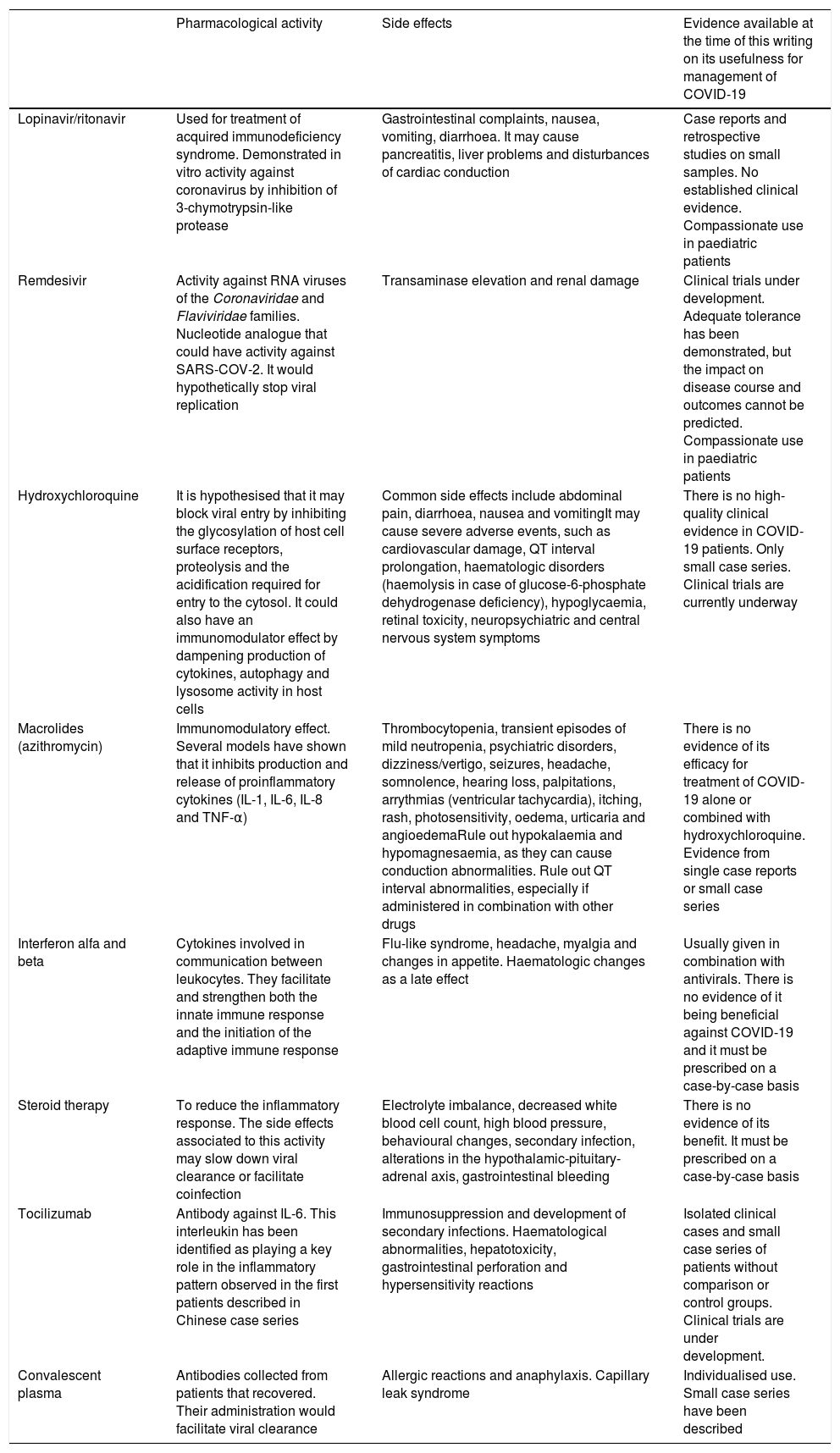
TreatmentsAt present, the treatment of COVID-19 mostly consists of symptom management and ventilatory support as needed.30 In some patients, such as those with risk factors or responding poorly, empiric treatment may be attempted with different drugs targeting either the virus or the dysregulated immune response. We now proceed to describe all the empiric treatments currently used.
Lopinavir/ritonavirThe use of both antivirals has been proposed for COVID-19 based on their previous performance against SARS-CoV and MERS-CoV. At present, there is no evidence supporting their use, and the early data on the subject suggest that they may not have an impact on mortality in these patients.31
RemdesivirThis drug has exhibited antiviral activity against a broad spectrum of RNA viruses, including SARS-CoV and MERS-CoV.6,9,32 Its effectiveness for treatment of Ebola is currently under investigation, as is its effectiveness and usefulness against SARS-CoV-2.32
HydroxychloroquineThe potential efficacy of hydroxychloroquine (HQ) against coronavirus was already described in the SARS-CoV epidemic. This molecule increases the pH of the environment, hindering the activity of the viral phagolysosome and therefore slowing down or inhibiting the replication of coronavirus. In vitro studies have recently demonstrated activity against SARS-CoV-2.33 There is no evidence from clinical trials supporting its efficacy in patients, and therefore it continues to be an empiric therapy.34 It is important to watch for the potential development of adverse effects when HQ is used either alone or in combination with other drugs.33,35
MacrolidesMacrolides have been shown to have certain immunomodulatory properties. Thus, they seem to have an effect on neutrophil activity or the release of cytokines such as IL-8. There is evidence that they can be useful for treatment of viral pneumonias and therefore have been proposed for adjuvant treatment in patients with COVID-19. There is still no evidence confirming that their use alone or in combination with HQ improves patient outcomes.35
Interferon alfa and betaThe main goal of administering interferon in the context of infection by coronavirus is to mitigate the potential disruption of this innate immune response pathway by the virus. This rationale was applied in previous epidemics by coronaviruses with controversial outcomes.
CorticosteroidsThe use of steroid therapy in patients with COVID-19 is controversial. On one hand, it may have an immunosuppressant effect that could be useful in case of proinflammatory immune dysregulation.29,36 On the other hand, these drugs exacerbate and prolong lymphopenia, and therefore could interfere in the response needed for viral clearance. In patients with respiratory illness caused by SARS-CoV, their use was not beneficial and was even associated with a longer duration of detectable viral loads (Table 1).
Description of the activity, side effects and evidence on the use of the main drugs proposed for treatment of COVID19. When it comes to the evidence, there are no published paediatric case series, so the evidence presented here refers to the adult population.
| Pharmacological activity | Side effects | Evidence available at the time of this writing on its usefulness for management of COVID-19 | |
|---|---|---|---|
| Lopinavir/ritonavir | Used for treatment of acquired immunodeficiency syndrome. Demonstrated in vitro activity against coronavirus by inhibition of 3-chymotrypsin-like protease | Gastrointestinal complaints, nausea, vomiting, diarrhoea. It may cause pancreatitis, liver problems and disturbances of cardiac conduction | Case reports and retrospective studies on small samples. No established clinical evidence. Compassionate use in paediatric patients |
| Remdesivir | Activity against RNA viruses of the Coronaviridae and Flaviviridae families. Nucleotide analogue that could have activity against SARS-COV-2. It would hypothetically stop viral replication | Transaminase elevation and renal damage | Clinical trials under development. Adequate tolerance has been demonstrated, but the impact on disease course and outcomes cannot be predicted. Compassionate use in paediatric patients |
| Hydroxychloroquine | It is hypothesised that it may block viral entry by inhibiting the glycosylation of host cell surface receptors, proteolysis and the acidification required for entry to the cytosol. It could also have an immunomodulator effect by dampening production of cytokines, autophagy and lysosome activity in host cells | Common side effects include abdominal pain, diarrhoea, nausea and vomitingIt may cause severe adverse events, such as cardiovascular damage, QT interval prolongation, haematologic disorders (haemolysis in case of glucose-6-phosphate dehydrogenase deficiency), hypoglycaemia, retinal toxicity, neuropsychiatric and central nervous system symptoms | There is no high-quality clinical evidence in COVID-19 patients. Only small case series. Clinical trials are currently underway |
| Macrolides (azithromycin) | Immunomodulatory effect. Several models have shown that it inhibits production and release of proinflammatory cytokines (IL-1, IL-6, IL-8 and TNF-α) | Thrombocytopenia, transient episodes of mild neutropenia, psychiatric disorders, dizziness/vertigo, seizures, headache, somnolence, hearing loss, palpitations, arrythmias (ventricular tachycardia), itching, rash, photosensitivity, oedema, urticaria and angioedemaRule out hypokalaemia and hypomagnesaemia, as they can cause conduction abnormalities. Rule out QT interval abnormalities, especially if administered in combination with other drugs | There is no evidence of its efficacy for treatment of COVID-19 alone or combined with hydroxychloroquine. Evidence from single case reports or small case series |
| Interferon alfa and beta | Cytokines involved in communication between leukocytes. They facilitate and strengthen both the innate immune response and the initiation of the adaptive immune response | Flu-like syndrome, headache, myalgia and changes in appetite. Haematologic changes as a late effect | Usually given in combination with antivirals. There is no evidence of it being beneficial against COVID-19 and it must be prescribed on a case-by-case basis |
| Steroid therapy | To reduce the inflammatory response. The side effects associated to this activity may slow down viral clearance or facilitate coinfection | Electrolyte imbalance, decreased white blood cell count, high blood pressure, behavioural changes, secondary infection, alterations in the hypothalamic-pituitary-adrenal axis, gastrointestinal bleeding | There is no evidence of its benefit. It must be prescribed on a case-by-case basis |
| Tocilizumab | Antibody against IL-6. This interleukin has been identified as playing a key role in the inflammatory pattern observed in the first patients described in Chinese case series | Immunosuppression and development of secondary infections. Haematological abnormalities, hepatotoxicity, gastrointestinal perforation and hypersensitivity reactions | Isolated clinical cases and small case series of patients without comparison or control groups. Clinical trials are under development. |
| Convalescent plasma | Antibodies collected from patients that recovered. Their administration would facilitate viral clearance | Allergic reactions and anaphylaxis. Capillary leak syndrome | Individualised use. Small case series have been described |
The recommendations made for steroid therapy by clinicians that managed patients with COVID-19 in Wuhan rested on 5 key points: (1) The risks and benefits of its use must be weighed carefully. (2) Its use should be considered mainly in critically ill patients. (3) Its use should be considered in patients with hypoxaemia due to underlying disease or previously receiving long-term steroid therapy. (4) It should be given at a low-to-medium dose (≤0.5–1mg/kg/day of methylprednisolone or equivalent agent), and (5) the duration of treatment should be short (≤7 days).37
TocilizumabAs discussed above, the synthesis of IL-6 by macrophages and T cells is part of the immune response against SARS-CoV-2. In cases of COVID-19 with a prolonged course or clinical worsening, there is frequently evidence of high serum levels of this cytokine. Tocilizumab is a humanised monoclonal antibody that inhibits ligand binding to the IL-6 receptor. It could be used to halt the underlying immune cell activation, and therefore could be a therapeutic option in patients with severe or progressing disease. If methods for quantification of this molecule were not available, ferritin could be used as a biomarker.12,18,30,36 At present, ferritin offers an adequate sensitivity and specificity to guide the diagnosis and followup of macrophage activation syndrome.28
Convalescent plasmaThe use of convalescent plasma was proposed in previous epidemics caused by SARS-CoV and MERS-CoV, and at present there are few data on its benefits. Only 1 study conducted in 5 patients suggested positive outcomes. Its use must be considered with caution, as it is not free of risks.38
In conclusion, SARS-CoV-2 may share some characteristics with previously known coronaviruses and have a similar effect on the immune response. Given its activity on the interferon signalling pathways and the way it dysregulates innate immunity, the use of treatments aimed at modulating or containing the innate immune response may be of interest. A better understanding of the underlying pathophysiological mechanisms or the identification of specific biomarkers (lymphocytes or IL-6 levels) could be useful in predicting the course of disease and developing individualised treatment plans with use of targeted therapies.
Conflicts of interestThe authors have no conflicts of interest to declare.
We thank all health care professionals for their contribution and help in this terrible situation. We also want to acknowledge patients and their families.
Please cite this article as: García-Salido A. Revisión narrativa sobre la respuesta inmunitaria frente a coronavirus: descripción general, aplicabilidad para SARS-COV-2 e implicaciones terapéuticas. An Pediatr (Barc). 2020;93:60.
