


Leptomeningeal dissemination in paediatric central nervous system (CNS) tumours is associated with a poor outcome, and new therapeutic strategies are desperately needed. One of the main difficulties in the treatment of CNS tumours is blood brain barrier penetration. Intrathecal therapy has shown to be effective in several paediatric tumours. The aim of this article is to review the data available on the use of liposomal cytarabine for paediatric patients with leptomeningeal dissemination of CNS tumours, including the pharmacology, administration, safety and efficacy data.
Los tumores pediátricos del sistema nervioso central (SNC) con diseminación leptomeníngea tienen mal pronóstico y es preciso encontrar nuevas alternativas terapéuticas. Una de las principales dificultades en el tratamiento de los tumores del SNC es la penetración de la barrera hematoencefálica, por lo que el tratamiento intratecal ha demostrado su eficacia en múltiples tumores pediátricos. En este artículo se revisa la experiencia disponible sobre la utilización de citarabina liposomal para pacientes pediátricos con tumores del SNC con diseminación leptomeníngea: farmacología, forma de administración, datos de seguridad y estudios de eficacia.
The meninges are the most frequent site of metastatic spread in paediatric central nervous system (CNS) tumours. This dissemination can be present at initial diagnosis or persist as minimal residual disease despite initial treatment and recur as metastatic disease.1 Another explanation for the phenomenon of persistent minimal residual disease and leptomeningeal metastatic recurrence is the presence of the blood–brain barrier (BBB), which blocks the delivery of drugs that are administered systemically.2
DiagnosisDissemination is suspected based on clinical symptoms and confirmed by abnormal MRI findings or the presence of cancerous cells in cerebrospinal fluid (CSF). The most frequent symptoms are headache, cranial nerve palsy, back pain or radiculalgia. Other possible manifestations include impaired CSF flow or reabsorption with secondary hydrocephalus. Diagnostic MRI findings can have a linear, nodular or mixed enhancement pattern. The diffuse pattern shows contrast uptake or thickening along the margins of the meninges, which can reach nerve root exits in the spinal region.3 Magnetic resonance abnormalities may be accompanied by the detection of malignant cells in CSF cytology, although in embryonal tumours it is possible to find evidence of dissemination in CSF cytology in the absence of abnormal MRI findings. The usefulness of flow cytometry of CSF samples from paediatric brain tumours has yet to be determined.
Incidence of metastatic dissemination in different types of paediatric tumoursThe highest incidence corresponds to embryonal tumours.4 In medulloblastoma, the most frequent type of malignant CSN tumour in the paediatric age group, the incidence of metastasis at the time of diagnosis ranges between 20% and 30%. Thirty percent of medulloblastomas do recur, usually with leptomeningeal dissemination. Metastatic disease becoming resistant to multimodal therapy is the usual cause of death in these patients, as opposed to local recurrence. The incidence of metastasis is particularly high in group 3, followed by group 4 and the Sonic Hedgehog medulloblastoma subgroups.5 In other embryonal tumours, such as the supratentorial primitive neuroectodermal tumours (PNET), which include pineoblastoma, embryonal tumours with multilayered rosettes (ETMRT), and atypical teratoid-rhabdoid tumours (ATRTs), dissemination may be found at the time of diagnosis or in a subsequent recurrence.
The incidence of dissemination is lower in gliomas.6,7 Recent studies on diffuse intrinsic pontine glioma revealed that up to 38% of patients had leptomeningeal spread at autopsy.8 A study on ependymoma reported that 9–20% of patients had dissemination at diagnosis, with a higher incidence in recurrent cases.9 Dissemination is rarely found in glioneural tumours, such as ganglioglioma or dysembryoplastic neuroectodermal tumour, but is a consistent finding in the rare cases of disseminated glioneural tumour.10 The incidence of dissemination in malignant germ cell tumours (germinomas, secretory germ cell tumours and mixed germ cell tumours) ranges between 10% and 20% at the time of diagnosis.11
The probability of developing CNS metastases from extracranial tumours is much lower in children and adolescents than in adults. The most frequent tumours with CNS metastases are sarcoma (Ewing sarcoma, osteosarcoma, synovial sarcoma), neuroblastoma, Wilms tumour and retinoblastoma.12
Treatment optionsThe goal of treatment for leptomeningeal dissemination can be preventive, when dissemination is not detectable by the usually available methods at initiation, or therapeutic, when there is evidence of metastatic disease in MRI or cytology. The treatment used most commonly is craniospinal irradiation covering the entire neural axis. In combination with chemotherapy, this approach has achieved cure rates in excess of 60% in patients with metastatic medulloblastoma, at the expense of mid- to long-term sequelae that are primarily neurocognitive. This limits its use in children aged less than 5 years.
Alternative treatment options are few and their mid- and long-term efficacy is poor. Most frequently, leptomeningeal dissemination is found after completion of multimodal frontline therapy, for instance in metastatic relapses of previously irradiated medulloblastoma. New therapies are needed to improve survival in refractory disease and reduce long-term sequelae in survivors. One possible strategy is the use of high doses of systemic chemotherapeutic agents such as methotrexate, or high doses of conventional chemotherapy followed by salvage therapy with hematopoietic stem cell transplantation, although this approach is not efficacious in previously irradiated relapsing medulloblastomas.13 When radiotherapy is not an option because it has been used previously or due to the age of the patient, one possible option to increase the penetrance of chemotherapy in the CNS is the direct injection of drugs via the intrathecal route.14 Few chemotherapeutic agents can be safely delivered intrathecally, and they have been studied more extensively in the context of leukaemias, although the use of other cytostatic agents (methotrexate, cytarabine, topotecan or etoposide) and new drugs (interferon alpha, interleukin 2 or anti-GD2 monoclonal antibodies) is currently being investigated. Intrathecal administration into the subarachnoid space via lumbar puncture has the advantage of bypassing the BBB. There are also drawbacks to repeated lumbar punctures with the corresponding anaesthesia and sedation: risk of inadequate injection technique, post-puncture headache, infection or haemorrhage. Placement of an Ommaya reservoir could alleviate some of these problems, allowing direct intraventricular delivery. Other drawbacks of intrathecal delivery are impaired CSF flow, CSF flow obstruction, and in patients with ventriculoperitoneal or ventriculoatrial shunts, inadequate drug exposure due to the shunt clearing the drug from the CSF space. Yet another drawback is their potential additive neurotoxic effect, of which there is evidence in drugs such as methotrexate.
In this article, we review the current evidence on the pharmacology, safety and efficacy of intrathecal liposomal cytarabine for the treatment of CNS tumours with leptomeningeal dissemination.
Pharmacology and pharmacokineticsCytarabine (Ara-C) is an antimetabolite, a pyrimidine nucleoside analogue of cytidine. It is converted intracellularly into the active metabolite arabinosylcytosine triphosphate (ara-CTP). It is a drug that acts specifically on the S phase of the cell cycle, inhibiting DNA synthesis (a competitive inhibitor of DNA polymerase).
Cytarabine is eliminated through its conversion to the inactive metabolite uracil arabinoside (ara-U) and subsequent secretion in the urine. When cytarabine is administered systemically it is quickly metabolised to ara-U, but in the CSF, conversion of cytarabine to ara-U is negligible due to the low activity of cytidine deaminase, so that the clearance rate of cytarabine in the CSF is similar to the rate of CSF bulk flow: 0.24mL per minute.15,16 Still, there is probably interindividual variability in the cytidine deaminase concentration in the brain and CSF, which seems to increase with age.16
The three anticancer drugs that are commonly administered via the intrathecal route are methotrexate, cytarabine and hydrocortisone. Both methotrexate and cytarabine act specifically on cells in the S phase of the cell cycle, and thus their efficacy increases with prolonged cytotoxic concentrations.15,16
The elimination half-life of free cytarabine following its intrathecal injection is 3.4h. Solid tumours have a lower proliferation rate than leukaemias and have higher cytotoxic thresholds that would require repeated intrathecal injections to maintain those concentrations in the CSF.15
Liposomal cytarabine is a cytarabine formulation that is prepared by packing an aqueous solution of the drug in capsules or multivesicular spherical particles15–18 that consist of vesicles made of concentric membranes, formed by one or more phospholipid bilayers, that hold an aqueous solution and allow the slow release of cytarabine to the CSF. Five hours after intrathecal administration, a peak in the concentration of free cytarabine is observed in the ventricular and lumbar sac regions. The exposure to the active principle in these two regions is similar, regardless of the route of administration.
The recommended dose of liposomal cytarabine for adults is 50mg delivered by intrathecal injection every two weeks, which results in an elimination half-life for free cytarabine in the CSF of 80h compared to the 3.4 half-life of non-liposomal cytarabine.17
Unlike changes in the body surface area, which continues to increase through childhood and adolescence, the volume of CSF, as happens with head circumference, increases rapidly in the first years of life and reaches its final volume at age 3–4 years.19 The doses of liposomal cytarabine tried out in children are based on this principle.
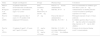
Table 1 summarises the currently available pharmacological data on the use of liposomal cytarabine in the paediatric population.
Studies on the pharmacology and pharmacokinetics of liposomal cytarabine in the paediatric population.
| Study | Sample size/diagnosis | Dosage | Pharmacology | Conclusion |
|---|---|---|---|---|
| Phase I trial: Bomgaars et al. (2004) | 19 patients with CNS involvement due to leukaemia, lymphoma or solid tumours (ages 3–21 years) | Dose escalation: 25mg followed by 35 and 50mg | Peak at 1–2h from administration Half-life, 50–57h | Dose recommended for children aged more than 3 years: 35mg Administration via lumbar puncture or reservoir achieves prolonged exposures of >7 days |
| Peyrl et al. (2009) | 4 children aged less than 3 years (11–35 months) with malignant CNS tumours | 25mg | Peak at the time of administration. Half-life, 59.3–56.7h | Prolonged exposure of up to 14 days achieved. In children aged less than 3 years, a 25mg dose achieved comparable drug exposure as 35mg in older children |
| Peyrl et al. (2014) | 15 patients (8 aged 3–10 years and 7 aged >10 years) | 35mg for ≤10 years 50mg for >10 years | Half-life, 31.5–40.9h in young children and 43.7–36.4h in children >10 years | Cytotoxic levels detected during week 1 in most patients |
Studies in children aged less than 3 years found levels similar to those found in older children, but lower than those found in adults.15,17,18 The concentrations of free cytarabine found in the intraventricular CSF following administration by lumbar puncture were comparable to those found following intraventricular administration, which demonstrates that cytarabine diffuses through the neural axis following lumbar injection. On the other hand, the concentrations of encapsulated cytarabine dropped faster following lumbar administration compared to cytarabine delivered intraventricularly. The authors concluded than in children aged less than 3 years, 25mg of intrathecal liposomal cytarabine can achieve the same drug exposure as doses of 35mg in children aged more than 3 years and doses of 50mg in adults, respectively.15
The same group of researchers studied the pharmacokinetics of intrathecal liposomal cytarabine administered every two to four weeks in doses of 35mg to children aged 3–10 years (8 patients) or 50mg in children aged more than 10 years (7 patients).16
These studies have demonstrated that liposomal preparations of cytarabine achieve prolonged cytotoxic concentrations following their intrathecal administration in children. The longer half-life observed in younger children could be due to lower concentrations of cytidine deaminase in the CSF of this subset of patients.
Paediatric dosage and administrationRecommended doses and administration regimensIn Spain, liposomal cytarabine has been approved for treatment of lymphomatous meningitis in adults. As is the case of other drugs commonly used in paediatrics that have not been properly tested in the paediatric population, there is some published evidence from several early clinical trials, pharmacokinetic studies and retrospective studies analysing clinical experience. Based on this evidence, there seems to be a consensus regarding the dosages that could be recommended in the paediatric age group. At any rate, such off-label use should always be approved following established procedure in each hospital, including the decision of a multidisciplinary board responsible for weighing the benefits and risks in each individual case, and the informed consent of the parents or legal guardians for the off-label use of the drug, which should include information on its expected benefits, its potential toxicities, and alternative treatments.
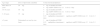
Table 2 presents the recommended doses per administration. It must be noted that the dose in children aged less than 3 years must be individualised, starting with a low dose, such as 1mg/kg, that can be increased to a maximum of 25mg in subsequent administrations.
Dosage and schedule of liposomal cytarabine treatment in paediatric patients.
| Age range | Dose of liposomal cytarabine | Corticosteroid prophylaxis |
|---|---|---|
| More than 18 years | 50mg | 4mg of intrathecal DXM 4mg oral/IV DXM every 12h for 2–3 day |
| 10–18 years | 50mg | 4mg of intrathecal DXM 4mg oral/IV DXM every 12h for 2–3 days |
| 3–10 years | 35mg | 2mg of intrathecal DXM 0.15mg/kg/dose (maximum 4mg/dose) oral/IV DXM every 12h for 2–3 days |
| <3 years | Determined on case-by-case basis | 1mg of intrathecal DXM 0.15mg/kg/dose (maximum 4mg/dose) oral/IV DXM every 12h for 2–3 days |
DXM, dexamethasone.
Several studies have shown that the use of corticosteroids decreases the incidence and severity of arachnoiditis associated with the use of liposomal cytarabine, with the best outcomes obtained with concomitant intrathecal administration of corticosteroids. Therefore, concomitant intrathecal administration of dexamethasone (see Table 2) is recommended with each dose. In some patients, dexamethasone may be substituted with intrathecal hydrocortisone, which is better tolerated.20 In addition, oral administration of dexamethasone is recommended three to five days after the administration of liposomal cytarabine, and the dose and regimen can be adjusted for each individual patient.
Mode of administrationLiposomal Cytarabine is administered by lumbar puncture or placement of an Ommaya reservoir. The procedure must be performed under deep sedation and analgesia, with one clinician performing sedation and a different clinician administering the drug. After collecting CSF, liposomal cytarabine must be infused slowly over one to five minutes, followed by the intrathecal administration of dexamethasone over one minute. If the drug is administered via lumbar puncture, the patient must remain in the supine position for at least an hour. The drugs must be administered by a clinician with demonstrated experience in lumbar puncture and the delivery of intrathecal chemotherapy.
Schedule. According to the summary of product characteristics, the first five doses must be administered every other week, followed by five more doses given every four weeks. No data are available on the safety of the continued use of liposomal cytarabine after 10 doses, and clinicians should weigh the risks and benefits of continuing treatment on a case-by-case basis.
Analysing CSF cytology is recommended at the time each dose is administered to monitor the cytologic response (in case the CSF had tested positive at baseline), as is performance of craniospinal MRI every eight to twelve weeks to assess the response of leptomeningeal dissemination.
ToxicityThe adverse reactions found most frequently in studies in adults are headache, arachnoiditis, pyrexia, weakness, nausea, vomiting, confusion, diarrhoea, thrombocytopaenia and fatigue. The main severe toxicities (in less than 10% of patients) are myelopathys, seizures, extreme somnolence, hemiplegia, visual changes, hearing loss and cranial nerve palsy. Other reported adverse effects include signs and symptoms of peripheral neuropathy such as pain, paraesthesias and even faecal and urinary incontinence.
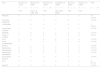
There are few data on the toxicity of cytarabine in children (see Table 3). In general, toxicity seems to be slightly lesser than the toxicity reported in adults. In the reviewed studies, forty-seven percent of children did not develop adverse reactions. The most common adverse reactions (in more than 5%) were headache, arachnoiditis, neurologic impairment, cauda equina syndrome, seizures, papilloedema and inflammation at the lumbar puncture site.
Toxicities associated most frequently with the use of liposomal cytarabine in paediatric patients.
| Type | Navajas et al. (2012) | Benesch et al. (2007) | Benesch et al. (2009) | Lassaletta et al. (2009) | Peyrl et al. (2009) | Sommer et al. (2008) | Total |
|---|---|---|---|---|---|---|---|
| N=11 | N=5 | N=19 | N=9 | N=6 | N=3 | N=53 | |
| CNS | R ALL, R AML, CNS | CNS | SNC, NHL, Rhabd | CNS | CNS | ||
| Headache | 5 | 0 | 0 | 1 | 1 | 3 | 10 (18.8%) |
| Neurologic impairment | 2 | 0 | 2 | 0 | 0 | 1 | 5 (9.4%) |
| Arachnoiditis | 1 | 0 | 4 | 2 | 0 | 2 | 9 (16.9%) |
| Cauda equina syndrome | 1 | 1 | 0 | 1 | 0 | 1 | 4 (7.5%) |
| Irritability | 1 | 0 | 0 | 1 | 0 | 0 | 2 (3.7%) |
| Nausea or vomiting | 2 | 0 | 0 | 0 | 0 | 0 | 2(3.7%) |
| Fever | 2 | 0 | 0 | 0 | 0 | 0 | 2 (3.7%) |
| Acute encephalopathy | 0 | 1 | 0 | 0 | 0 | 0 | 1 (1.8%) |
| Seizures | 1 | 1 | 0 | 1 | 0 | 0 | 3 (5.6%) |
| Papilloedema | 0 | 0 | 0 | 0 | 0 | 3 | 3 (5.6%) |
| Radiculopathy | 0 | 0 | 0 | 0 | 0 | 1 | 1 (1.8%) |
| Post-puncture inflammation | 0 | 0 | 0 | 0 | 0 | 2 | 2 (3.7%) |
| Post-puncture infection | 0 | 0 | 0 | 0 | 0 | 1 | 1 (1.8%) |
| None | 3 | 2 | 11 | 4 | 5 | 0 | 25 (47.1%) |
| Grade IV | 0 | 0 | 2 | 0 | 0 | 0 | 2 (4.2%) |
Liposomal cytarabine is one of the drugs that has been studied most extensively in the past decade for the treatment or prophylaxis of leptomeningeal dissemination via intrathecal administration. At the time of this writing, there are no randomised studies comparing the efficacy of liposomal cytarabine to that of other drugs in the paediatric population.
As is often the case in paediatric diseases, data from studies in adults are used to assess its potential use in this age group. There is evidence of a cytologic response in adults with leptomeningeal dissemination and different histological types, such as lung cancer, melanoma, PNET and medulloblastoma, among others.21
Studies in adults comparing it with intrathecal methotrexate seem to show a better response to liposomal cytarabine, although this trend was not statistically significant. However, significant differences have been found in the median time to neurologic progression (30 days for methotrexate versus 58 days for liposomal cytarabine).22
All the evidence on its use in brain tumours in the paediatric age group comes from single-arm studies, many of which do not differentiate between de novo and relapsed patients, or between its therapeutic and prophylaxis use, making it difficult to draw solid conclusions.
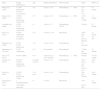
Approximately 100 cases of children and adolescents with CNS tumours treated with liposomal cytarabine have been described in the literature (see Table 4), often in combination with other treatment modalities, most of which were cases of relapsing embryonal cell tumours. Cytologic responses have been described in the treatment of leptomeningeal dissemination with an acceptable toxicity profile, but the data did not suffice to clearly prove that this cytologic efficacy correlates with an improvement in survival.
Data on the efficacy of liposomal cytarabine in paediatric patients with CNS tumours.
| Study | Sample size/diagnosis | Age | Intrathecal treatment | Other treatment | Status | Follow up |
|---|---|---|---|---|---|---|
| Partap et al. (2010) | 17 CNS (embryonal) 5 de novo 12 recurrences | <30 | 6 doses (1–16) | Chemotherapy+RTX | 5/17 alive 4/5 1/12 | 48 months |
| Benesch et al. (2009) | 19 CNS 14 CNS (embryonal) 5 other CNS | <23 | 4 doses (1–10) | Chemotherapy+RTX | 13/19 alive 3 in FR | 12 months |
| Lassaletta et al. (2009) | 9 CNS 5 CNS (embryonal) 4 other CNS | <4 | 4 doses (1–7) | Chemotherapy 2 RTX | 5/9 alive | 20 months |
| Peyrl et al. (2012) | 16 CNS (embryonal) 5 recurrent no Depocyt | 0–12 | 10 doses (1–18) | Metronomic | 10/16 alive 7/11 alive on Depocyt | 33 months (10–58) |
| Bomgaars et al. (2004) | 7 CNS 6 CNS (embryonal) | 3–21 | 3 doses (2–10) | Chemotherapy | 7/7 deceased | |
| Mastronuzzi et al. (2013) | 3 CNS (embryonal) | 6–17 | Not specified | Chemotherapy | 2/3 alive and diseased | 10 months |
| Slavc et al. (2014) | 8 CNS (ATRT) | 0–17 | 5 doses (LC in only 5 patients) | Chemotherapy+RTX | 8/8 alive | 16–197 months |
| Navajas et al. (2012) | 20 CNS 15 CNS (embryonal) 5 Other CNS | 8 months–18 years | 5 doses (1–9) | Chemotherapy | 10/18 alive (2 n.d.) 6/13 alive (2 n.d.) | >180 days |
| Peyrl et al. (2014) | 16 CNS 9 CNS (embryonal) 7 other | 3–18 years | 6 doses (3–18) | Chemotherapy | 8/16 alive 6/9 alive | 6–69 months |
| Nygaard et al. (2011) | 1 (medulloblastoma) | 14 | 13 doses | Metronomic | Alive | 34 months |
| Total | 116 CNS/94 embryonal | 60/114 alive | ||||
CNS, central nervous system; FR, full remission; LC, liposomal cytarabine; n.d., not documented; RTX, radiotherapy.
A nationwide, multicentre study conducted in Spain included 20 cases of CNS tumours with leptomeningeal dissemination: four ependymomas, 10 medulloblastomas, three supratentorial PNETs and three other cases, which included both de novo and relapse cases.23 Approximately, half of the patients received concomitant chemotherapy without radiotherapy. Eleven out of nineteen patients (59%) responded to treatment, with five achieving complete remission and six partial remission. The response was neurologic in eleven out of nineteen and cytologic in seven out of ten of the evaluable cases, with a median progression-free survival of 180 days and development of adverse effects in 11 out of 20 cases.
Another case series presented a retrospective study of patients aged less than 30 years with CNS embryonal tumours: five de novo cases (3 ATRT, 2 of supratentorial PNET) and twelve recurrent cases (all medulloblastoma).24 None of the patients with negative CSF cytology at diagnosis developed malignant CSF cytology during treatment. Six patients with malignant cytology treated with at least two doses (mean, 3 doses) cleared their CSF. The survival in this study was four out of five in de novo patients and one out of twelve in relapsed patients (median, 26.8 months). Yet another published series reported favourable outcomes of its use with concomitant antiangiogenic therapy in patients with medulloblastoma. When it comes to ATRT, a study with a small sample of patients has reported promising results.25 A phase II clinical trial is needed to assess the efficacy of liposomal cytarabine in embryonal CNS tumours.
The data currently available is insufficient to provide recommendations supported by high quality evidence. Nevertheless, patients with CNS tumours with leptomeningeal dissemination have a very poor prognosis, and in many cases there are no potentially curative therapeutic alternatives for them. Therefore, the use of liposomal cytarabine could be considered in various situations:
- –
1st or subsequent recurrences of supratentorial PNET or medulloblastoma with leptomeningeal dissemination.
- –
1st or subsequent recurrences of ependymoma with leptomeningeal dissemination.
- –
In patients with ATRT at the time of diagnosis and until initiation of radiotherapy.
- –
In patients with leptomeningeal dissemination of any other type of CNS tumour, excluding gliomas, until initiation of radiotherapy, or in patients that are ineligible for radiotherapy (for example, children aged less than 3 years).
Paediatric embryonal CNS tumours with leptomeningeal dissemination continue to pose significant challenges to the paediatric oncologist due to a dearth of therapeutic options both in relapse patients and for cases that carry a poor prognosis from the time of diagnosis. Treatment with intrathecal liposomal cytarabine is an alternative to consider in selected cases. The toxicity profile is acceptable when cytarabine is combined with prophylactic treatment with intrathecal and oral dexamethasone and pharmacotherapy is performed in facilities with experience in the treatment of CNS tumours. Although the potential long-term applications of liposomal cytarabine remain to be determined by large-scope studies, the current literature reports favourable cytologic and clinical outcomes in a proportion of patients with high-risk tumours that have experienced multiple relapses.
Conflict of interestsAll authors of this article have participated in an advisory board on the use of liposomal cytarabine in paediatric patients.
Please cite this article as: Moreno L, García Ariza MA, Cruz O, Calvo C, Fuster JL, Salinas JA, et al. Citarabina liposomal para el tratamiento de la diseminación leptomeníngea en tumores del sistema nervioso central en niños y adolescentes. An Pediatr (Barc). 2016;85:274.
