


A boy aged 16 months presented in the emergency department with asthenia, abdominal pain and vomiting of 72 h’ duration. The key finding of the physical examination was the detection on palpation of an abdominal mass in the umbilical region.
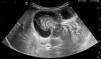
The mass was evaluated with an abdominal ultrasound scan (Fig. 1), leading to diagnosis of advanced ileocecal intussusception and possible bowel perforation.

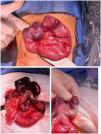
The patient underwent urgent surgery via midline laparotomy, which revealed ileocecal intussusception extending to the hepatic flexure (Fig. 2A).

Reduction of the intussusception made visible a necrotic ileal segment with a round, indurated lesion that acted as the head of intussusception (Figs. 2B and C). The affected bowel was resected, followed by end-to-end anastomosis, and the surgical specimen submitted for pathological examination.
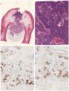
The histological study confirmed the presence of heterotropic pancreas at the level of the ileum (Fig. 3), which had caused the intussusception.

Histological study.
Top left: transmural lesion extending from the mucosa to the serous layer, composed of pancreatic ducts and acini but without islets of Langerhans. Top right: at greater magnification, pancreatic acini could be visualized, composed of polygonal cells without atypia. Bottom: immunohistochemical staining was positive for neuroendocrine markers such as chromogranin (left) and synaptophysin (right).
The patient did not experience any postoperative complications.
Heterotopic or ectopic pancreas is defined as the presence of pancreatic tissue outside the normal anatomical location of the pancreas.1 It is a rare condition in the pediatric population that, while usually asymptomatic, may manifest in the form of pancreatitis, infection, malignancy or intussusception (with heterotropic pancreas accounting for 1%–2% of total cases of intussusception).2
