array:24 [
  "pii" => "S2341287923002259"
  "issn" => "23412879"
  "doi" => "10.1016/j.anpede.2023.10.001"
  "estado" => "S300"
  "fechaPublicacion" => "2023-11-01"
  "aid" => "3434"
  "copyright" => "Asociación Española de Pediatría"
  "copyrightAnyo" => "2023"
  "documento" => "article"
  "crossmark" => 1
  "subdocumento" => "sco"
  "cita" => "An Pediatr (Barc). 2023;99:360-1"
  "abierto" => array:3 [
    "ES" => true
    "ES2" => true
    "LATM" => true
  ]
  "gratuito" => true
  "lecturas" => array:1 [
    "total" => 0
  ]
  "Traduccion" => array:1 [
    "es" => array:19 [
      "pii" => "S1695403323000784"
      "issn" => "16954033"
      "doi" => "10.1016/j.anpedi.2023.03.009"
      "estado" => "S300"
      "fechaPublicacion" => "2023-11-01"
      "aid" => "3434"
      "copyright" => "Asociación Española de Pediatría"
      "documento" => "article"
      "crossmark" => 1
      "subdocumento" => "sco"
      "cita" => "An Pediatr (Barc). 2023;99:360-1"
      "abierto" => array:3 [
        "ES" => true
        "ES2" => true
        "LATM" => true
      ]
      "gratuito" => true
      "lecturas" => array:1 [
        "total" => 0
      ]
      "es" => array:11 [
        "idiomaDefecto" => true
        "cabecera" => "<span class="elsevierStyleTextfn">Im&#225;genes en pediatr&#237;a</span>"
        "titulo" => "Ictiosis como manifestaci&#243;n de enfermedad de Gaucher fetal"
        "tienePdf" => "es"
        "tieneTextoCompleto" => "es"
        "paginas" => array:1 [
          0 => array:2 [
            "paginaInicial" => "360"
            "paginaFinal" => "361"
          ]
        ]
        "titulosAlternativos" => array:1 [
          "en" => array:1 [
            "titulo" => "Ichtyosis as a manifestation of fetal Gaucher"
          ]
        ]
        "contieneTextoCompleto" => array:1 [
          "es" => true
        ]
        "contienePdf" => array:1 [
          "es" => true
        ]
        "resumenGrafico" => array:2 [
          "original" => 0
          "multimedia" => array:7 [
            "identificador" => "fig0010"
            "etiqueta" => "Figura 2"
            "tipo" => "MULTIMEDIAFIGURA"
            "mostrarFloat" => true
            "mostrarDisplay" => false
            "figura" => array:1 [
              0 => array:4 [
                "imagen" => "gr2.jpeg"
                "Alto" => 476
                "Ancho" => 905
                "Tamanyo" => 75061
              ]
            ]
            "descripcion" => array:1 [
              "es" => "<p id="spar0010" class="elsevierStyleSimplePara elsevierViewall">Al sexto d&#237;a de vida</p>"
            ]
          ]
        ]
        "autores" => array:1 [
          0 => array:2 [
            "autoresLista" => "Francisco A&#46; Palaz&#243;n-Rico, Jose Mar&#237;a Lloreda-Garc&#237;a"
            "autores" => array:2 [
              0 => array:2 [
                "nombre" => "Francisco A&#46;"
                "apellidos" => "Palaz&#243;n-Rico"
              ]
              1 => array:2 [
                "nombre" => "Jose Mar&#237;a"
                "apellidos" => "Lloreda-Garc&#237;a"
              ]
            ]
          ]
        ]
      ]
      "idiomaDefecto" => "es"
      "Traduccion" => array:1 [
        "en" => array:9 [
          "pii" => "S2341287923002259"
          "doi" => "10.1016/j.anpede.2023.10.001"
          "estado" => "S300"
          "subdocumento" => ""
          "abierto" => array:3 [
            "ES" => true
            "ES2" => true
            "LATM" => true
          ]
          "gratuito" => true
          "lecturas" => array:1 [
            "total" => 0
          ]
          "idiomaDefecto" => "en"
          "EPUB" => "https://multimedia.elsevier.es/PublicationsMultimediaV1/item/epub/S2341287923002259?idApp=UINPBA00005H"
        ]
      ]
      "EPUB" => "https://multimedia.elsevier.es/PublicationsMultimediaV1/item/epub/S1695403323000784?idApp=UINPBA00005H"
      "url" => "/16954033/0000009900000005/v2_202311111135/S1695403323000784/v2_202311111135/es/main.assets"
    ]
  ]
  "itemSiguiente" => array:19 [
    "pii" => "S2341287923002302"
    "issn" => "23412879"
    "doi" => "10.1016/j.anpede.2023.10.004"
    "estado" => "S300"
    "fechaPublicacion" => "2023-11-01"
    "aid" => "3451"
    "copyright" => "Asociaci&#243;n Espa&#241;ola de Pediatr&#237;a"
    "documento" => "article"
    "crossmark" => 1
    "subdocumento" => "sco"
    "cita" => "An Pediatr &#40;Barc&#41;. 2023;99:362-3"
    "abierto" => array:3 [
      "ES" => true
      "ES2" => true
      "LATM" => true
    ]
    "gratuito" => true
    "lecturas" => array:1 [
      "total" => 0
    ]
    "en" => array:11 [
      "idiomaDefecto" => true
      "cabecera" => "<span class="elsevierStyleTextfn">Images in Paediatrics</span>"
      "titulo" => "Self-healing juvenile cutaneous mucinosis"
      "tienePdf" => "en"
      "tieneTextoCompleto" => "en"
      "paginas" => array:1 [
        0 => array:2 [
          "paginaInicial" => "362"
          "paginaFinal" => "363"
        ]
      ]
      "titulosAlternativos" => array:1 [
        "es" => array:1 [
          "titulo" => "Mucinosis cut&#225;nea juvenil autorresolutiva"
        ]
      ]
      "contieneTextoCompleto" => array:1 [
        "en" => true
      ]
      "contienePdf" => array:1 [
        "en" => true
      ]
      "resumenGrafico" => array:2 [
        "original" => 0
        "multimedia" => array:8 [
          "identificador" => "fig0015"
          "etiqueta" => "Figure 3"
          "tipo" => "MULTIMEDIAFIGURA"
          "mostrarFloat" => true
          "mostrarDisplay" => false
          "figura" => array:1 [
            0 => array:4 [
              "imagen" => "gr3.jpeg"
              "Alto" => 1179
              "Ancho" => 755
              "Tamanyo" => 150104
            ]
          ]
          "detalles" => array:1 [
            0 => array:3 [
              "identificador" => "at0045"
              "detalle" => "Figure "
              "rol" => "short"
            ]
          ]
          "descripcion" => array:1 [
            "en" => "<p id="spar0015" class="elsevierStyleSimplePara elsevierViewall">Biopsy sample of a cutaneous lesion in the patient&#46; Alcian blue stain evincing mucin deposition&#46;</p>"
          ]
        ]
      ]
      "autores" => array:1 [
        0 => array:2 [
          "autoresLista" => "Elvira Reneses Prieto, Agust&#237;n L&#243;pez L&#243;pez, Elena M&#46;&#170; L&#243;pez-Negrete Arenal, Inmaculada Olmedo D&#237;az"
          "autores" => array:4 [
            0 => array:2 [
              "nombre" => "Elvira"
              "apellidos" => "Reneses Prieto"
            ]
            1 => array:2 [
              "nombre" => "Agust&#237;n"
              "apellidos" => "L&#243;pez L&#243;pez"
            ]
            2 => array:2 [
              "nombre" => "Elena M&#46;&#170;"
              "apellidos" => "L&#243;pez-Negrete Arenal"
            ]
            3 => array:2 [
              "nombre" => "Inmaculada"
              "apellidos" => "Olmedo D&#237;az"
            ]
          ]
        ]
      ]
    ]
    "idiomaDefecto" => "en"
    "Traduccion" => array:1 [
      "es" => array:9 [
        "pii" => "S1695403323001121"
        "doi" => "10.1016/j.anpedi.2023.05.001"
        "estado" => "S300"
        "subdocumento" => ""
        "abierto" => array:3 [
          "ES" => true
          "ES2" => true
          "LATM" => true
        ]
        "gratuito" => true
        "lecturas" => array:1 [
          "total" => 0
        ]
        "idiomaDefecto" => "es"
        "EPUB" => "https://multimedia.elsevier.es/PublicationsMultimediaV1/item/epub/S1695403323001121?idApp=UINPBA00005H"
      ]
    ]
    "EPUB" => "https://multimedia.elsevier.es/PublicationsMultimediaV1/item/epub/S2341287923002302?idApp=UINPBA00005H"
    "url" => "/23412879/0000009900000005/v1_202311240521/S2341287923002302/v1_202311240521/en/main.assets"
  ]
  "itemAnterior" => array:19 [
    "pii" => "S2341287923002247"
    "issn" => "23412879"
    "doi" => "10.1016/j.anpede.2023.09.010"
    "estado" => "S300"
    "fechaPublicacion" => "2023-11-01"
    "aid" => "3433"
    "copyright" => "Asociaci&#243;n Espa&#241;ola de Pediatr&#237;a"
    "documento" => "article"
    "crossmark" => 1
    "subdocumento" => "sco"
    "cita" => "An Pediatr &#40;Barc&#41;. 2023;99:358-9"
    "abierto" => array:3 [
      "ES" => true
      "ES2" => true
      "LATM" => true
    ]
    "gratuito" => true
    "lecturas" => array:1 [
      "total" => 0
    ]
    "en" => array:11 [
      "idiomaDefecto" => true
      "cabecera" => "<span class="elsevierStyleTextfn">Images in Paediatrics</span>"
      "titulo" => "Lymphangioma of the palatine tonsil"
      "tienePdf" => "en"
      "tieneTextoCompleto" => "en"
      "paginas" => array:1 [
        0 => array:2 [
          "paginaInicial" => "358"
          "paginaFinal" => "359"
        ]
      ]
      "titulosAlternativos" => array:1 [
        "es" => array:1 [
          "titulo" => "Linfangioma de am&#237;gdala palatina"
        ]
      ]
      "contieneTextoCompleto" => array:1 [
        "en" => true
      ]
      "contienePdf" => array:1 [
        "en" => true
      ]
      "resumenGrafico" => array:2 [
        "original" => 0
        "multimedia" => array:8 [
          "identificador" => "fig0005"
          "etiqueta" => "Figure 1"
          "tipo" => "MULTIMEDIAFIGURA"
          "mostrarFloat" => true
          "mostrarDisplay" => false
          "figura" => array:1 [
            0 => array:4 [
              "imagen" => "gr1.jpeg"
              "Alto" => 480
              "Ancho" => 755
              "Tamanyo" => 50545
            ]
          ]
          "detalles" => array:1 [
            0 => array:3 [
              "identificador" => "at0005"
              "detalle" => "Figure "
              "rol" => "short"
            ]
          ]
          "descripcion" => array:1 [
            "en" => "<p id="spar0005" class="elsevierStyleSimplePara elsevierViewall">&#40;A&#41; Right tonsil measuring 3&#8239;&#215;&#8239;2&#8239;cm with a polypoid overgrowth measuring 1&#46;5&#8239;cm &#40;length&#41;&#8239;&#215;&#8239;1&#8239;cm &#40;thickness&#41;&#46; &#40;B&#41; Left tonsil with normal morphology measuring 2&#46;5&#8239;&#215;&#8239;2&#8239;cm&#46;</p>"
          ]
        ]
      ]
      "autores" => array:1 [
        0 => array:2 [
          "autoresLista" => "Giselle Cuestas, Mariela Anah&#237; Maidana Roa, Mar&#237;a Teresa Garc&#237;a de D&#225;vila"
          "autores" => array:3 [
            0 => array:2 [
              "nombre" => "Giselle"
              "apellidos" => "Cuestas"
            ]
            1 => array:2 [
              "nombre" => "Mariela Anah&#237;"
              "apellidos" => "Maidana Roa"
            ]
            2 => array:2 [
              "nombre" => "Mar&#237;a Teresa"
              "apellidos" => "Garc&#237;a de D&#225;vila"
            ]
          ]
        ]
      ]
    ]
    "idiomaDefecto" => "en"
    "Traduccion" => array:1 [
      "es" => array:9 [
        "pii" => "S1695403323000772"
        "doi" => "10.1016/j.anpedi.2023.03.008"
        "estado" => "S300"
        "subdocumento" => ""
        "abierto" => array:3 [
          "ES" => true
          "ES2" => true
          "LATM" => true
        ]
        "gratuito" => true
        "lecturas" => array:1 [
          "total" => 0
        ]
        "idiomaDefecto" => "es"
        "EPUB" => "https://multimedia.elsevier.es/PublicationsMultimediaV1/item/epub/S1695403323000772?idApp=UINPBA00005H"
      ]
    ]
    "EPUB" => "https://multimedia.elsevier.es/PublicationsMultimediaV1/item/epub/S2341287923002247?idApp=UINPBA00005H"
    "url" => "/23412879/0000009900000005/v1_202311240521/S2341287923002247/v1_202311240521/en/main.assets"
  ]
  "en" => array:15 [
    "idiomaDefecto" => true
    "cabecera" => "<span class="elsevierStyleTextfn">Images in Paediatrics</span>"
    "titulo" => "Ichthyosis as a manifestation of fetal gaucher"
    "tieneTextoCompleto" => true
    "paginas" => array:1 [
      0 => array:2 [
        "paginaInicial" => "360"
        "paginaFinal" => "361"
      ]
    ]
    "autores" => array:1 [
      0 => array:4 [
        "autoresLista" => "Francisco A&#46; Palaz&#243;n-Rico, Jose Mar&#237;a Lloreda-Garc&#237;a"
        "autores" => array:2 [
          0 => array:4 [
            "nombre" => "Francisco A&#46;"
            "apellidos" => "Palaz&#243;n-Rico"
            "email" => array:1 [
              0 => "palaxote@gmail.com"
            ]
            "referencia" => array:2 [
              0 => array:2 [
                "etiqueta" => "<span class="elsevierStyleSup">a</span>"
                "identificador" => "aff0005"
              ]
              1 => array:2 [
                "etiqueta" => "<span class="elsevierStyleSup">&#42;</span>"
                "identificador" => "cor0005"
              ]
            ]
          ]
          1 => array:3 [
            "nombre" => "Jose Mar&#237;a"
            "apellidos" => "Lloreda-Garc&#237;a"
            "referencia" => array:1 [
              0 => array:2 [
                "etiqueta" => "<span class="elsevierStyleSup">b</span>"
                "identificador" => "aff0010"
              ]
            ]
          ]
        ]
        "afiliaciones" => array:2 [
          0 => array:3 [
            "entidad" => "Servicio de Pediatr&#237;a&#44; Hospital Universitario Santa Luc&#237;a&#44; Cartagena &#40;Murcia&#41;&#44; Spain"
            "etiqueta" => "a"
            "identificador" => "aff0005"
          ]
          1 => array:3 [
            "entidad" => "Unidad de Neonatolog&#237;a&#44; Hospital Universitario Santa Luc&#237;a&#44; Cartagena &#40;Murcia&#41;&#44; Spain"
            "etiqueta" => "b"
            "identificador" => "aff0010"
          ]
        ]
        "correspondencia" => array:1 [
          0 => array:3 [
            "identificador" => "cor0005"
            "etiqueta" => "&#8270;"
            "correspondencia" => "Corresponding author&#46;"
          ]
        ]
      ]
    ]
    "titulosAlternativos" => array:1 [
      "es" => array:1 [
        "titulo" => "Ictiosis como manifestaci&#243;n de enfermedad de gaucher fetal"
      ]
    ]
    "resumenGrafico" => array:2 [
      "original" => 0
      "multimedia" => array:8 [
        "identificador" => "fig0015"
        "etiqueta" => "Figure 3"
        "tipo" => "MULTIMEDIAFIGURA"
        "mostrarFloat" => true
        "mostrarDisplay" => false
        "figura" => array:1 [
          0 => array:4 [
            "imagen" => "gr3.jpeg"
            "Alto" => 1037
            "Ancho" => 755
            "Tamanyo" => 55134
          ]
        ]
        "detalles" => array:1 [
          0 => array:3 [
            "identificador" => "at0015"
            "detalle" => "Figure "
            "rol" => "short"
          ]
        ]
        "descripcion" => array:1 [
          "en" => "<p id="spar0015" class="elsevierStyleSimplePara elsevierViewall">Hepatosplenomegaly&#46;</p>"
        ]
      ]
    ]
    "textoCompleto" => "<span class="elsevierStyleSections"><p id="par0005" class="elsevierStylePara elsevierViewall">We present the case of a male term neonate with no family history of interest&#44; who had an uncomplicated birth and an Apgar score of 9&#8211;10&#46; He was the second-born child of nonconsanguineous parents&#46; There was no previous history of miscarriage or abnormalities in prenatal care tests&#46;</p><p id="par0010" class="elsevierStylePara elsevierViewall">The salient findings of the physical examination were a dry&#44; erythematous skin with desquamation and diffuse petechiae &#40;<a class="elsevierStyleCrossRefs" href="#fig0005">Figs&#46; 1 and 2</a>&#41; associated with hypotrophic appearance&#44; jaundice and weak sucking&#46;</p><elsevierMultimedia ident="fig0005"></elsevierMultimedia><elsevierMultimedia ident="fig0010"></elsevierMultimedia><p id="par0015" class="elsevierStylePara elsevierViewall">The patient was admitted to the intensive care unit&#44; with subsequent detection of coagulopathy&#44; cholestasis and persistent thrombocytopenia despite multiple transfusions&#46; The results of cultures and serological tests were negative&#46; The peripheral blood smear examination revealed the presence of vacuolated lymphocytes&#46; The postnatal radiograph and ultrasound examinations detected hepatosplenomegaly &#40;<a class="elsevierStyleCrossRef" href="#fig0015">Fig&#46; 3</a>&#41;&#46;</p><elsevierMultimedia ident="fig0015"></elsevierMultimedia><p id="par0020" class="elsevierStylePara elsevierViewall">Ichthyosis associated with a genetic disorder was suspected&#44; and the differential diagnosis included foetal Gaucher disease&#44; Chanarin-Dorfman syndrome and NISCH syndrome&#46; The enzyme test for Gaucher disease ordered on day 8 post birth evinced the absence of glucocerebrosidase&#46; The evaluation continued with an ichthyosis panel&#44; adding molecular tests for Gaucher disease and NISCH syndrome&#46; This resulted in the detection of two heterozygous variants of the <span class="elsevierStyleItalic">GBA</span> gene &#40;p&#46;Pro430Leu and p&#46;Leu483Pro&#44; which were present in the parents&#41;&#46; The patient died 2 months later during a respiratory infection&#46;</p><p id="par0025" class="elsevierStylePara elsevierViewall">Foetal Gaucher disease<a class="elsevierStyleCrossRef" href="#bib0005"><span class="elsevierStyleSup">1</span></a> has an incidence of less than 1 case per 10<span class="elsevierStyleSup">6</span> live births and a prevalence of nearly zero on account of the lack of effective treatment&#44; early mortality and forms of disease leading to miscarriage or hydrops fetalis&#46; In the rest of cases&#44; it is associated with hepatosplenomegaly and sustained cytopenia and may present as an ichthyosiform syndrome&#46;<a class="elsevierStyleCrossRef" href="#bib0010"><span class="elsevierStyleSup">2</span></a></p><p id="par0030" class="elsevierStylePara elsevierViewall">It is caused by changes in the <span class="elsevierStyleItalic">GBA</span> gene &#40;1q21&#41;&#44; resulting in glucocerebrosidase deficiency&#46; Forms associated with the p&#46;Leu483Pro variant have a poorer prognosis&#46;<a class="elsevierStyleCrossRef" href="#bib0015"><span class="elsevierStyleSup">3</span></a></p><p id="par0035" class="elsevierStylePara elsevierViewall">Awareness of the association between thrombocytopenia and ichthyosis is important in order to diagnose affected patients and provide genetic counselling to families&#46;</p></span>"
    "pdfFichero" => "main.pdf"
    "tienePdf" => true
    "fechaRecibido" => "2023-01-20"
    "fechaAceptado" => "2023-03-22"
    "multimedia" => array:3 [
      0 => array:8 [
        "identificador" => "fig0005"
        "etiqueta" => "Figure 1"
        "tipo" => "MULTIMEDIAFIGURA"
        "mostrarFloat" => true
        "mostrarDisplay" => false
        "figura" => array:1 [
          0 => array:4 [
            "imagen" => "gr1.jpeg"
            "Alto" => 1005
            "Ancho" => 755
            "Tamanyo" => 113118
          ]
        ]
        "detalles" => array:1 [
          0 => array:3 [
            "identificador" => "at0005"
            "detalle" => "Figure "
            "rol" => "short"
          ]
        ]
        "descripcion" => array:1 [
          "en" => "<p id="spar0005" class="elsevierStyleSimplePara elsevierViewall">Appearance at birth&#46;</p>"
        ]
      ]
      1 => array:8 [
        "identificador" => "fig0010"
        "etiqueta" => "Figure 2"
        "tipo" => "MULTIMEDIAFIGURA"
        "mostrarFloat" => true
        "mostrarDisplay" => false
        "figura" => array:1 [
          0 => array:4 [
            "imagen" => "gr2.jpeg"
            "Alto" => 476
            "Ancho" => 905
            "Tamanyo" => 75061
          ]
        ]
        "detalles" => array:1 [
          0 => array:3 [
            "identificador" => "at0010"
            "detalle" => "Figure "
            "rol" => "short"
          ]
        ]
        "descripcion" => array:1 [
          "en" => "<p id="spar0010" class="elsevierStyleSimplePara elsevierViewall">Day 6 post birth&#46;</p>"
        ]
      ]
      2 => array:8 [
        "identificador" => "fig0015"
        "etiqueta" => "Figure 3"
        "tipo" => "MULTIMEDIAFIGURA"
        "mostrarFloat" => true
        "mostrarDisplay" => false
        "figura" => array:1 [
          0 => array:4 [
            "imagen" => "gr3.jpeg"
            "Alto" => 1037
            "Ancho" => 755
            "Tamanyo" => 55134
          ]
        ]
        "detalles" => array:1 [
          0 => array:3 [
            "identificador" => "at0015"
            "detalle" => "Figure "
            "rol" => "short"
          ]
        ]
        "descripcion" => array:1 [
          "en" => "<p id="spar0015" class="elsevierStyleSimplePara elsevierViewall">Hepatosplenomegaly&#46;</p>"
        ]
      ]
    ]
    "bibliografia" => array:2 [
      "titulo" => "References"
      "seccion" => array:1 [
        0 => array:2 [
          "identificador" => "bibs0005"
          "bibliografiaReferencia" => array:3 [
            0 => array:3 [
              "identificador" => "bib0005"
              "etiqueta" => "1"
              "referencia" => array:1 [
                0 => array:2 [
                  "contribucion" => array:1 [
                    0 => array:2 [
                      "titulo" => "Gaucher Disease"
                      "autores" => array:1 [
                        0 => array:2 [
                          "etal" => false
                          "autores" => array:2 [
                            0 => "G&#46;M&#46; Pastores"
                            1 => "D&#46;A&#46; Hughes"
                          ]
                        ]
                      ]
                    ]
                  ]
                  "host" => array:2 [
                    0 => array:1 [
                      "LibroEditado" => array:5 [
                        "editores" => "M&#46;P&#46;Adam, D&#46;B&#46;Everman, G&#46;M&#46;Mirzaa"
                        "titulo" => "GeneReviews&#174; &#91;Internet&#93;"
                        "paginaInicial" => "1993"
                        "paginaFinal" => "2022"
                        "serieFecha" => "2000"
                      ]
                    ]
                    1 => array:1 [
                      "WWW" => array:1 [
                        "link" => "https&#58;&#47;&#47;www&#46;ncbi&#46;nlm&#46;nih&#46;gov&#47;books&#47;NBK1269&#47;"
                      ]
                    ]
                  ]
                ]
              ]
            ]
            1 => array:3 [
              "identificador" => "bib0010"
              "etiqueta" => "2"
              "referencia" => array:1 [
                0 => array:2 [
                  "contribucion" => array:1 [
                    0 => array:2 [
                      "titulo" => "Inherited ichthyoses&#47;generalized Mendelian disorders of cornification"
                      "autores" => array:1 [
                        0 => array:2 [
                          "etal" => true
                          "autores" => array:6 [
                            0 => "M&#46; Schmuth"
                            1 => "V&#46; Martinz"
                            2 => "A&#46;R&#46; Janecke"
                            3 => "C&#46; Fauth"
                            4 => "A&#46; Schossig"
                            5 => "J&#46; Zschocke"
                          ]
                        ]
                      ]
                    ]
                  ]
                  "host" => array:1 [
                    0 => array:2 [
                      "doi" => "10.1038/ejhg.2012.121"
                      "Revista" => array:5 [
                        "tituloSerie" => "Eur J Hum Genet"
                        "fecha" => "2013"
                        "volumen" => "21"
                        "paginaInicial" => "123"
                        "paginaFinal" => "133"
                      ]
                    ]
                  ]
                ]
              ]
            ]
            2 => array:3 [
              "identificador" => "bib0015"
              "etiqueta" => "3"
              "referencia" => array:1 [
                0 => array:2 [
                  "contribucion" => array:1 [
                    0 => array:2 [
                      "titulo" => "A review of gaucher disease pathophysiology&#44; clinical presentation and treatments"
                      "autores" => array:1 [
                        0 => array:2 [
                          "etal" => true
                          "autores" => array:6 [
                            0 => "J&#46; Stirnemann"
                            1 => "N&#46; Belmatoug"
                            2 => "F&#46; Camou"
                            3 => "C&#46; Serratrice"
                            4 => "R&#46; Froissart"
                            5 => "C&#46; Caillaud"
                          ]
                        ]
                      ]
                    ]
                  ]
                  "host" => array:1 [
                    0 => array:2 [
                      "doi" => "10.3390/ijms18020441"
                      "Revista" => array:6 [
                        "tituloSerie" => "Int J Mol Sci &#91;Internet&#93;"
                        "fecha" => "2017"
                        "volumen" => "18"
                        "numero" => "2"
                        "paginaInicial" => "441"
                        "link" => array:1 [
                          0 => array:2 [
                            "url" => "https://www.ncbi.nlm.nih.gov/pubmed/28218669"
                            "web" => "Medline"
                          ]
                        ]
                      ]
                    ]
                  ]
                ]
              ]
            ]
          ]
        ]
      ]
    ]
  ]
  "idiomaDefecto" => "en"
  "url" => "/23412879/0000009900000005/v1_202311240521/S2341287923002259/v1_202311240521/en/main.assets"
  "Apartado" => array:4 [
    "identificador" => "77701"
    "tipo" => "SECCION"
    "en" => array:2 [
      "titulo" => "Images in Paediatrics"
      "idiomaDefecto" => true
    ]
    "idiomaDefecto" => "en"
  ]
  "PDF" => "https://static.elsevier.es/multimedia/23412879/0000009900000005/v1_202311240521/S2341287923002259/v1_202311240521/en/main.pdf?idApp=UINPBA00005H&text.app=https://analesdepediatria.org/"
  "EPUB" => "https://multimedia.elsevier.es/PublicationsMultimediaV1/item/epub/S2341287923002259?idApp=UINPBA00005H"
]
Share
Journal Information
Vol. 99. Issue 5.
Pages 360-361 (1 November 2023)
Vol. 99. Issue 5.
Pages 360-361 (1 November 2023)
Images in Paediatrics
Full text access
Ichthyosis as a manifestation of fetal gaucher
Ictiosis como manifestación de enfermedad de gaucher fetal
Visits
1649
Francisco A. Palazón-Ricoa,
Corresponding author
palaxote@gmail.com

Corresponding author.
, Jose María Lloreda-Garcíab
a Servicio de Pediatría, Hospital Universitario Santa Lucía, Cartagena (Murcia), Spain
b Unidad de Neonatología, Hospital Universitario Santa Lucía, Cartagena (Murcia), Spain
This item has received
Article information
Full Text
Bibliography
Download PDF
Statistics
Figures (3)
Show moreShow less
Full Text

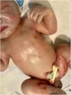
We present the case of a male term neonate with no family history of interest, who had an uncomplicated birth and an Apgar score of 9–10. He was the second-born child of nonconsanguineous parents. There was no previous history of miscarriage or abnormalities in prenatal care tests.

The salient findings of the physical examination were a dry, erythematous skin with desquamation and diffuse petechiae (Figs. 1 and 2) associated with hypotrophic appearance, jaundice and weak sucking.

Figure 1.

Appearance at birth.

(0.11MB).
Figure 2.

Day 6 post birth.

(0.07MB).

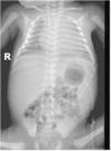
The patient was admitted to the intensive care unit, with subsequent detection of coagulopathy, cholestasis and persistent thrombocytopenia despite multiple transfusions. The results of cultures and serological tests were negative. The peripheral blood smear examination revealed the presence of vacuolated lymphocytes. The postnatal radiograph and ultrasound examinations detected hepatosplenomegaly (Fig. 3).

Figure 3.

Hepatosplenomegaly.

(0.05MB).

Ichthyosis associated with a genetic disorder was suspected, and the differential diagnosis included foetal Gaucher disease, Chanarin-Dorfman syndrome and NISCH syndrome. The enzyme test for Gaucher disease ordered on day 8 post birth evinced the absence of glucocerebrosidase. The evaluation continued with an ichthyosis panel, adding molecular tests for Gaucher disease and NISCH syndrome. This resulted in the detection of two heterozygous variants of the GBA gene (p.Pro430Leu and p.Leu483Pro, which were present in the parents). The patient died 2 months later during a respiratory infection.

Foetal Gaucher disease1 has an incidence of less than 1 case per 106 live births and a prevalence of nearly zero on account of the lack of effective treatment, early mortality and forms of disease leading to miscarriage or hydrops fetalis. In the rest of cases, it is associated with hepatosplenomegaly and sustained cytopenia and may present as an ichthyosiform syndrome.2

It is caused by changes in the GBA gene (1q21), resulting in glucocerebrosidase deficiency. Forms associated with the p.Leu483Pro variant have a poorer prognosis.3

Awareness of the association between thrombocytopenia and ichthyosis is important in order to diagnose affected patients and provide genetic counselling to families.

References
[1]
G.M. Pastores, D.A. Hughes.
Gaucher Disease.
GeneReviews® [Internet], pp. 1993-2022
[2]
M. Schmuth, V. Martinz, A.R. Janecke, C. Fauth, A. Schossig, J. Zschocke, et al.
Inherited ichthyoses/generalized Mendelian disorders of cornification.
Eur J Hum Genet, 21 (2013), pp. 123-133
[3]
J. Stirnemann, N. Belmatoug, F. Camou, C. Serratrice, R. Froissart, C. Caillaud, et al.
A review of gaucher disease pathophysiology, clinical presentation and treatments.
Int J Mol Sci [Internet], 18 (2017), pp. 441
Copyright © 2023. Asociación Española de Pediatría
Download PDF
Idiomas
Anales de Pediatría (English Edition)
Article options
Tools
es en

¿Es usted profesional sanitario apto para prescribir o dispensar medicamentos?

Are you a health professional able to prescribe or dispense drugs?