A patient aged 13 years presented with a mass in the right popliteal fossa detected the previous day. It was not preceded by trauma, infectious disease or any other relevant history. In the examination palpation, the mass was pulsatile and painless on palpation, the pedal pulse was preserved and there were no signs of venous thrombosis or distal ischaemia. The patient also exhibited mild scoliosis and joint hypermobility, with no other signs of connective tissue disease and no muscle involvement.
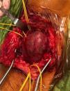
The findings of an ultrasound scan (Fig. 1) and a CT angiogram (Fig. 2) led to diagnosis of giant popliteal aneurysm. The echocardiogram and ophthalmological evaluation were normal. The patient was classified as high priority to underwent early surgical intervention (Fig. 3), consisting of reversed saphenous vein grafting.
Genetic testing detected a mutation in the COL12A1 gene (variant c.5839C>A p.Pro1947Thr), which, while classified as a variant of uncertain significance, involves a gene previously associated with connective tissue and muscular disease.
Popliteal aneurysm is infrequent in childhood. It is more frequent in the context of collagen diseases, although it is very rarely the initial symptom of these diseases.1 It is usually asymptomatic, but may manifest with thrombosis or ruptured aneurysm, with a high risk for ischaemia in the affected extremity.2,3 The treatment is based on the complete isolation of the aneurysm by percutaneous stenting or surgical bypass, the latter of which is preferred in children and patients with collagen diseases.3
FundingThis research did not receive any external funding.
Conflicts of interestThe authors have no conflicts of interest to declare.


