array:23 [
  "pii" => "S2341287924000218"
  "issn" => "23412879"
  "doi" => "10.1016/j.anpede.2024.01.012"
  "estado" => "S300"
  "fechaPublicacion" => "2024-03-01"
  "aid" => "3562"
  "copyright" => "Asociación Española de Pediatría"
  "copyrightAnyo" => "2024"
  "documento" => "article"
  "crossmark" => 1
  "subdocumento" => "sco"
  "cita" => "An Pediatr (Barc). 2024;100:228-9"
  "abierto" => array:3 [
    "ES" => true
    "ES2" => true
    "LATM" => true
  ]
  "gratuito" => true
  "lecturas" => array:1 [
    "total" => 0
  ]
  "Traduccion" => array:1 [
    "es" => array:19 [
      "pii" => "S1695403324000018"
      "issn" => "16954033"
      "doi" => "10.1016/j.anpedi.2023.11.013"
      "estado" => "S300"
      "fechaPublicacion" => "2024-03-01"
      "aid" => "3562"
      "copyright" => "Asociación Española de Pediatría"
      "documento" => "article"
      "crossmark" => 1
      "subdocumento" => "sco"
      "cita" => "An Pediatr (Barc). 2024;100:228-9"
      "abierto" => array:3 [
        "ES" => true
        "ES2" => true
        "LATM" => true
      ]
      "gratuito" => true
      "lecturas" => array:1 [
        "total" => 0
      ]
      "es" => array:11 [
        "idiomaDefecto" => true
        "cabecera" => "<span class="elsevierStyleTextfn">Im&#225;genes en pediatr&#237;a</span>"
        "titulo" => "Quiste de la bolsa de Blake&#58; diagn&#243;stico mediante imagen por resonancia magn&#233;tica"
        "tienePdf" => "es"
        "tieneTextoCompleto" => "es"
        "paginas" => array:1 [
          0 => array:2 [
            "paginaInicial" => "228"
            "paginaFinal" => "229"
          ]
        ]
        "titulosAlternativos" => array:1 [
          "en" => array:1 [
            "titulo" => "Blake&#39;s pouch cyst&#58; Diagnosis by Magnetic Resonance Imaging"
          ]
        ]
        "contieneTextoCompleto" => array:1 [
          "es" => true
        ]
        "contienePdf" => array:1 [
          "es" => true
        ]
        "resumenGrafico" => array:2 [
          "original" => 0
          "multimedia" => array:7 [
            "identificador" => "fig0005"
            "etiqueta" => "Figura 1"
            "tipo" => "MULTIMEDIAFIGURA"
            "mostrarFloat" => true
            "mostrarDisplay" => false
            "figura" => array:1 [
              0 => array:4 [
                "imagen" => "gr1.jpeg"
                "Alto" => 600
                "Ancho" => 1674
                "Tamanyo" => 117053
              ]
            ]
            "descripcion" => array:1 [
              "es" => "<p id="spar0005" class="elsevierStyleSimplePara elsevierViewall">Resonancia magn&#233;tica preoperatoria en lactante var&#243;n de 6 meses con hidroc&#233;falo tetraventricular grave&#46; &#40;A&#41; Plano sagital en el que se observa la apertura del acueducto cerebral &#40;punta de flecha blanca&#41; entre el cuarto ventr&#237;culo dilatado y el quiste de la bolsa de Blake &#40;estrella negra&#41;&#46; Pared posterior fina del quiste &#40;flecha blanca&#41; inferior a los l&#243;bulos ligeramente elevados y comprimidos del vermis cerebeloso posterior&#46; Concavidad del suelo del tercer ventr&#237;culo como consecuencia de la acumulaci&#243;n de l&#237;quido cefalorraqu&#237;deo ventricular al nivel de las cisternas &#40;flecha negra&#41;&#46; &#40;B&#41; Imagen axial de los agujeros de Luschka&#44; que aparec&#237;an abiertos &#40;flechas blancas&#41;&#46; &#40;C&#41; Imagen axial que muestra hidrocefalia asociada a edema intersticial periventricular &#40;flecha blanca&#41; indicativo de aumento de la presi&#243;n intracraneal al nivel del septo pel&#250;cido perforado &#40;flecha negra&#41;&#46;</p>"
            ]
          ]
        ]
        "autores" => array:1 [
          0 => array:2 [
            "autoresLista" => "Gabriel Miura, Deborah Desiree Coelho Marra, M&#225;rcio Lu&#237;s Duarte, Marcelo de Queiroz Pereira da Silva"
            "autores" => array:4 [
              0 => array:2 [
                "nombre" => "Gabriel"
                "apellidos" => "Miura"
              ]
              1 => array:2 [
                "nombre" => "Deborah Desiree Coelho"
                "apellidos" => "Marra"
              ]
              2 => array:2 [
                "nombre" => "M&#225;rcio Lu&#237;s"
                "apellidos" => "Duarte"
              ]
              3 => array:2 [
                "nombre" => "Marcelo de Queiroz Pereira da"
                "apellidos" => "Silva"
              ]
            ]
          ]
        ]
      ]
      "idiomaDefecto" => "es"
      "Traduccion" => array:1 [
        "en" => array:9 [
          "pii" => "S2341287924000218"
          "doi" => "10.1016/j.anpede.2024.01.012"
          "estado" => "S300"
          "subdocumento" => ""
          "abierto" => array:3 [
            "ES" => true
            "ES2" => true
            "LATM" => true
          ]
          "gratuito" => true
          "lecturas" => array:1 [
            "total" => 0
          ]
          "idiomaDefecto" => "en"
          "EPUB" => "https://multimedia.elsevier.es/PublicationsMultimediaV1/item/epub/S2341287924000218?idApp=UINPBA00005H"
        ]
      ]
      "EPUB" => "https://multimedia.elsevier.es/PublicationsMultimediaV1/item/epub/S1695403324000018?idApp=UINPBA00005H"
      "url" => "/16954033/0000010000000003/v1_202403130517/S1695403324000018/v1_202403130517/es/main.assets"
    ]
  ]
  "itemAnterior" => array:19 [
    "pii" => "S2341287924000322"
    "issn" => "23412879"
    "doi" => "10.1016/j.anpede.2023.12.015"
    "estado" => "S300"
    "fechaPublicacion" => "2024-03-01"
    "aid" => "3561"
    "copyright" => "Asociaci&#243;n Espa&#241;ola de Pediatr&#237;a"
    "documento" => "article"
    "crossmark" => 1
    "subdocumento" => "sco"
    "cita" => "An Pediatr &#40;Barc&#41;. 2024;100:226-7"
    "abierto" => array:3 [
      "ES" => true
      "ES2" => true
      "LATM" => true
    ]
    "gratuito" => true
    "lecturas" => array:1 [
      "total" => 0
    ]
    "en" => array:11 [
      "idiomaDefecto" => true
      "cabecera" => "<span class="elsevierStyleTextfn">Images in Paediatrics</span>"
      "titulo" => "Bipartite sesamoid in adolescent"
      "tienePdf" => "en"
      "tieneTextoCompleto" => "en"
      "paginas" => array:1 [
        0 => array:2 [
          "paginaInicial" => "226"
          "paginaFinal" => "227"
        ]
      ]
      "titulosAlternativos" => array:1 [
        "es" => array:1 [
          "titulo" => "Sesamoideo bipartito en adolescente"
        ]
      ]
      "contieneTextoCompleto" => array:1 [
        "en" => true
      ]
      "contienePdf" => array:1 [
        "en" => true
      ]
      "resumenGrafico" => array:2 [
        "original" => 0
        "multimedia" => array:8 [
          "identificador" => "fig0005"
          "etiqueta" => "Figure 1"
          "tipo" => "MULTIMEDIAFIGURA"
          "mostrarFloat" => true
          "mostrarDisplay" => false
          "figura" => array:1 [
            0 => array:4 [
              "imagen" => "gr1.jpeg"
              "Alto" => 1089
              "Ancho" => 755
              "Tamanyo" => 74312
            ]
          ]
          "detalles" => array:1 [
            0 => array:3 [
              "identificador" => "at0005"
              "detalle" => "Figure "
              "rol" => "short"
            ]
          ]
          "descripcion" => array:1 [
            "en" => "<p id="spar0005" class="elsevierStyleSimplePara elsevierViewall">Radiograph of the right foot&#46; Bipartite sesamoid&#46;</p>"
          ]
        ]
      ]
      "autores" => array:1 [
        0 => array:2 [
          "autoresLista" => "Beatriz Jim&#233;nez de la Jara, Jana Izquierdo Frechilla, Elena Davydoff Pastor, Miguel &#193;ngel Molina Guti&#233;rrez"
          "autores" => array:4 [
            0 => array:2 [
              "nombre" => "Beatriz"
              "apellidos" => "Jim&#233;nez de la Jara"
            ]
            1 => array:2 [
              "nombre" => "Jana"
              "apellidos" => "Izquierdo Frechilla"
            ]
            2 => array:2 [
              "nombre" => "Elena"
              "apellidos" => "Davydoff Pastor"
            ]
            3 => array:2 [
              "nombre" => "Miguel &#193;ngel"
              "apellidos" => "Molina Guti&#233;rrez"
            ]
          ]
        ]
      ]
    ]
    "idiomaDefecto" => "en"
    "Traduccion" => array:1 [
      "es" => array:9 [
        "pii" => "S1695403323002734"
        "doi" => "10.1016/j.anpedi.2023.12.010"
        "estado" => "S300"
        "subdocumento" => ""
        "abierto" => array:3 [
          "ES" => true
          "ES2" => true
          "LATM" => true
        ]
        "gratuito" => true
        "lecturas" => array:1 [
          "total" => 0
        ]
        "idiomaDefecto" => "es"
        "EPUB" => "https://multimedia.elsevier.es/PublicationsMultimediaV1/item/epub/S1695403323002734?idApp=UINPBA00005H"
      ]
    ]
    "EPUB" => "https://multimedia.elsevier.es/PublicationsMultimediaV1/item/epub/S2341287924000322?idApp=UINPBA00005H"
    "url" => "/23412879/0000010000000003/v1_202403190604/S2341287924000322/v1_202403190604/en/main.assets"
  ]
  "en" => array:16 [
    "idiomaDefecto" => true
    "cabecera" => "<span class="elsevierStyleTextfn">Images in Paediatrics</span>"
    "titulo" => "Blake&#39;s pouch cyst&#58; diagnosis by MRI"
    "tieneTextoCompleto" => true
    "paginas" => array:1 [
      0 => array:2 [
        "paginaInicial" => "228"
        "paginaFinal" => "229"
      ]
    ]
    "autores" => array:1 [
      0 => array:4 [
        "autoresLista" => "Gabriel Miura, Deborah Desiree Coelho Marra, M&#225;rcio Lu&#237;s Duarte, Marcelo de Queiroz Pereira da Silva"
        "autores" => array:4 [
          0 => array:3 [
            "nombre" => "Gabriel"
            "apellidos" => "Miura"
            "referencia" => array:1 [
              0 => array:2 [
                "etiqueta" => "<span class="elsevierStyleSup">a</span>"
                "identificador" => "aff0005"
              ]
            ]
          ]
          1 => array:3 [
            "nombre" => "Deborah Desiree Coelho"
            "apellidos" => "Marra"
            "referencia" => array:1 [
              0 => array:2 [
                "etiqueta" => "<span class="elsevierStyleSup">a</span>"
                "identificador" => "aff0005"
              ]
            ]
          ]
          2 => array:4 [
            "nombre" => "M&#225;rcio Lu&#237;s"
            "apellidos" => "Duarte"
            "email" => array:1 [
              0 => "mlduarte@unaerp.br"
            ]
            "referencia" => array:2 [
              0 => array:2 [
                "etiqueta" => "<span class="elsevierStyleSup">b</span>"
                "identificador" => "aff0010"
              ]
              1 => array:2 [
                "etiqueta" => "<span class="elsevierStyleSup">&#42;</span>"
                "identificador" => "cor0005"
              ]
            ]
          ]
          3 => array:3 [
            "nombre" => "Marcelo de Queiroz Pereira da"
            "apellidos" => "Silva"
            "referencia" => array:1 [
              0 => array:2 [
                "etiqueta" => "<span class="elsevierStyleSup">c</span>"
                "identificador" => "aff0015"
              ]
            ]
          ]
        ]
        "afiliaciones" => array:3 [
          0 => array:3 [
            "entidad" => "Faculdade de Ci&#234;ncias M&#233;dicas de Santos&#44; Santos&#44; SP&#44; Brasil"
            "etiqueta" => "a"
            "identificador" => "aff0005"
          ]
          1 => array:3 [
            "entidad" => "Universidade de Ribeir&#227;o Preto Campus Guaruj&#225;&#44; Guaruj&#225;&#44; SP&#44; Brasil"
            "etiqueta" => "b"
            "identificador" => "aff0010"
          ]
          2 => array:3 [
            "entidad" => "Medical of WebImagem&#44; S&#227;o Paulo&#44; SP&#44; Brasil"
            "etiqueta" => "c"
            "identificador" => "aff0015"
          ]
        ]
        "correspondencia" => array:1 [
          0 => array:3 [
            "identificador" => "cor0005"
            "etiqueta" => "&#8270;"
            "correspondencia" => "Corresponding author&#46;"
          ]
        ]
      ]
    ]
    "titulosAlternativos" => array:1 [
      "es" => array:1 [
        "titulo" => "Quiste de la bolsa de Blake&#58; diagn&#243;stico mediante RM"
      ]
    ]
    "resumenGrafico" => array:2 [
      "original" => 0
      "multimedia" => array:8 [
        "identificador" => "fig0005"
        "etiqueta" => "Figure 1"
        "tipo" => "MULTIMEDIAFIGURA"
        "mostrarFloat" => true
        "mostrarDisplay" => false
        "figura" => array:1 [
          0 => array:4 [
            "imagen" => "gr1.jpeg"
            "Alto" => 600
            "Ancho" => 1674
            "Tamanyo" => 117053
          ]
        ]
        "detalles" => array:1 [
          0 => array:3 [
            "identificador" => "at0005"
            "detalle" => "Figure "
            "rol" => "short"
          ]
        ]
        "descripcion" => array:1 [
          "en" => "<p id="spar0005" class="elsevierStyleSimplePara elsevierViewall">Preoperative MRI in a male infant aged 6 months with severe tetraventricular hydrocephalus&#46; &#40;A&#41; Sagittal image evincing patency of the cerebral aqueduct &#40;white arrowhead&#41; between the enlarged fourth ventricle and Blake&#8217;s pouch cyst &#40;black star&#41;&#46; Thin posterior cyst wall &#40;white arrow&#41; inferior to the slightly elevated and compressed lobules of the posterior vermis&#46; Concavity of the floor of the third ventricle as a consequence of the cerebrospinal fluid outflow from the ventricles into the cisterns &#40;black arrow&#41;&#46; &#40;B&#41; Axial MRI evincing patency of the foramina of Luschka &#40;white arrows&#41;&#46; &#40;C&#41; Axial image showing hydrocephalus associated with periventricular interstitial oedema &#40;white arrow&#41; indicative of increased intracranial pressure at the level of the perforated septum pellucidum &#40;black arrow&#41;&#46;</p>"
        ]
      ]
    ]
    "textoCompleto" => "<span class="elsevierStyleSections"><p id="par0005" class="elsevierStylePara elsevierViewall">Blake&#39;s pouch cyst is defined as a midline cystic malformation consisting of an posterior outpouching of the inferior medullary velum into the cisterna magna&#46;<a class="elsevierStyleCrossRef" href="#bib0005"><span class="elsevierStyleSup">1</span></a> It is attributed to the lack of fenestration of Blake&#8217;s Pouch&#44; an embryological expansion of the posterior membranous area continuous with the fourth ventricle&#44; which would normally give rise to the foramen of Magendie&#46;<a class="elsevierStyleCrossRef" href="#bib0010"><span class="elsevierStyleSup">2</span></a> Failed fenestration results in the lack of communication between the fourth ventricle and the subarachnoid space&#44; which in turn can cause hydrocephalus&#46;<a class="elsevierStyleCrossRef" href="#bib0010"><span class="elsevierStyleSup">2</span></a> The differential diagnosis includes arachnoid cyst&#44; mega cisterna magna and Dandy-Walker variant&#46;<a class="elsevierStyleCrossRef" href="#bib0010"><span class="elsevierStyleSup">2</span></a></p><p id="par0010" class="elsevierStylePara elsevierViewall">Fenestration of the cyst is one the approaches proposed to treat the main complication of Blake&#8217;s pouch cyst&#44; hydrocephalus&#46; In children aged less than 1 year&#44; fenestration is often unsuccessful and the patient requires placement of a ventriculoperitoneal shunt&#46;<a class="elsevierStyleCrossRefs" href="#bib0005"><span class="elsevierStyleSup">1&#44;3</span></a> Endoscopic third ventriculostomy is the treatment of choice&#46;<a class="elsevierStyleCrossRef" href="#bib0015"><span class="elsevierStyleSup">3</span></a></p><p id="par0015" class="elsevierStylePara elsevierViewall">We present the case of a male infant aged 6 months assessed for macrocephaly of 2 month&#8217;s duration&#44; born at 38 weeks of gestation and with no history of gestational or perinatal complications&#46; The physical examination revealed that the infant was not able to support his head or roll&#44; milestones that the mother reported he had achieved before the onset of the condition with hypoactivity and vomiting&#46; She also denied any convulsive seizures since birth&#46; Magnetic resonance imaging &#40;MRI&#41; allowed visualization of Blake&#8217;s pouch cyst associated with signs of intracranial hypertension &#40;<a class="elsevierStyleCrossRef" href="#fig0005">Fig&#46; 1</a>&#41;&#46; We referred the patient for placement of a ventriculoperitoneal shunt&#46;</p><elsevierMultimedia ident="fig0005"></elsevierMultimedia><span id="sec0040" class="elsevierStyleSection elsevierViewall"><span class="elsevierStyleSectionTitle" id="sect0100">Financial Disclosure</span><p id="par0180" class="elsevierStylePara elsevierViewall">Authors state that no economic support has been associated with the present study&#46;</p></span><span id="sec1040" class="elsevierStyleSection elsevierViewall"><span class="elsevierStyleSectionTitle" id="sect1100">Conflicts of Interest</span><p id="par1180" class="elsevierStylePara elsevierViewall">Authors declare no conflict of interest regarding the present study&#46;</p></span></span>"
    "textoCompletoSecciones" => array:1 [
      "secciones" => array:3 [
        0 => array:2 [
          "identificador" => "sec0040"
          "titulo" => "Financial Disclosure"
        ]
        1 => array:2 [
          "identificador" => "sec1040"
          "titulo" => "Conflicts of Interest"
        ]
        2 => array:1 [
          "titulo" => "References"
        ]
      ]
    ]
    "pdfFichero" => "main.pdf"
    "tienePdf" => true
    "fechaRecibido" => "2023-07-05"
    "fechaAceptado" => "2023-11-15"
    "multimedia" => array:1 [
      0 => array:8 [
        "identificador" => "fig0005"
        "etiqueta" => "Figure 1"
        "tipo" => "MULTIMEDIAFIGURA"
        "mostrarFloat" => true
        "mostrarDisplay" => false
        "figura" => array:1 [
          0 => array:4 [
            "imagen" => "gr1.jpeg"
            "Alto" => 600
            "Ancho" => 1674
            "Tamanyo" => 117053
          ]
        ]
        "detalles" => array:1 [
          0 => array:3 [
            "identificador" => "at0005"
            "detalle" => "Figure "
            "rol" => "short"
          ]
        ]
        "descripcion" => array:1 [
          "en" => "<p id="spar0005" class="elsevierStyleSimplePara elsevierViewall">Preoperative MRI in a male infant aged 6 months with severe tetraventricular hydrocephalus&#46; &#40;A&#41; Sagittal image evincing patency of the cerebral aqueduct &#40;white arrowhead&#41; between the enlarged fourth ventricle and Blake&#8217;s pouch cyst &#40;black star&#41;&#46; Thin posterior cyst wall &#40;white arrow&#41; inferior to the slightly elevated and compressed lobules of the posterior vermis&#46; Concavity of the floor of the third ventricle as a consequence of the cerebrospinal fluid outflow from the ventricles into the cisterns &#40;black arrow&#41;&#46; &#40;B&#41; Axial MRI evincing patency of the foramina of Luschka &#40;white arrows&#41;&#46; &#40;C&#41; Axial image showing hydrocephalus associated with periventricular interstitial oedema &#40;white arrow&#41; indicative of increased intracranial pressure at the level of the perforated septum pellucidum &#40;black arrow&#41;&#46;</p>"
        ]
      ]
    ]
    "bibliografia" => array:2 [
      "titulo" => "References"
      "seccion" => array:1 [
        0 => array:2 [
          "identificador" => "bibs0005"
          "bibliografiaReferencia" => array:3 [
            0 => array:3 [
              "identificador" => "bib0005"
              "etiqueta" => "1"
              "referencia" => array:1 [
                0 => array:2 [
                  "contribucion" => array:1 [
                    0 => array:2 [
                      "titulo" => "Endoscopic third ventriculostomy for the treatment of Blake&#8217;s pouch cyst in adulthood"
                      "autores" => array:1 [
                        0 => array:2 [
                          "etal" => false
                          "autores" => array:5 [
                            0 => "L&#46; Mongardi"
                            1 => "M&#46; D&#8217;Andrea"
                            2 => "L&#46; Tosatto"
                            3 => "M&#46; Volpin"
                            4 => "F&#46; Cultrera"
                          ]
                        ]
                      ]
                    ]
                  ]
                  "host" => array:1 [
                    0 => array:2 [
                      "doi" => "10.1007/s13760-020-01591-0"
                      "Revista" => array:7 [
                        "tituloSerie" => "Acta Neurol Belg&#46;"
                        "fecha" => "2022"
                        "volumen" => "122"
                        "numero" => "2"
                        "paginaInicial" => "537"
                        "paginaFinal" => "539"
                        "link" => array:1 [
                          0 => array:2 [
                            "url" => "https://www.ncbi.nlm.nih.gov/pubmed/33420942"
                            "web" => "Medline"
                          ]
                        ]
                      ]
                    ]
                  ]
                ]
              ]
            ]
            1 => array:3 [
              "identificador" => "bib0010"
              "etiqueta" => "2"
              "referencia" => array:1 [
                0 => array:2 [
                  "contribucion" => array:1 [
                    0 => array:2 [
                      "titulo" => "Blake&#39;s pouch cysts and differential diagnoses in prenatal and postnatal MRI &#58; a pictorial review"
                      "autores" => array:1 [
                        0 => array:2 [
                          "etal" => true
                          "autores" => array:6 [
                            0 => "T&#46; Kau"
                            1 => "R&#46; Marterer"
                            2 => "R&#46; Kottke"
                            3 => "R&#46; Birnbacher"
                            4 => "J&#46; Gellen"
                            5 => "E&#46; Nagy"
                          ]
                        ]
                      ]
                    ]
                  ]
                  "host" => array:1 [
                    0 => array:2 [
                      "doi" => "10.1007/s00062-019-00871-4"
                      "Revista" => array:7 [
                        "tituloSerie" => "Clin Neuroradiol"
                        "fecha" => "2020"
                        "volumen" => "30"
                        "numero" => "3"
                        "paginaInicial" => "435"
                        "paginaFinal" => "445"
                        "link" => array:1 [
                          0 => array:2 [
                            "url" => "https://www.ncbi.nlm.nih.gov/pubmed/31942658"
                            "web" => "Medline"
                          ]
                        ]
                      ]
                    ]
                  ]
                ]
              ]
            ]
            2 => array:3 [
              "identificador" => "bib0015"
              "etiqueta" => "3"
              "referencia" => array:1 [
                0 => array:2 [
                  "contribucion" => array:1 [
                    0 => array:2 [
                      "titulo" => "Blake&#8217;s pouch cyst&#58; prenatal diagnosis and management"
                      "autores" => array:1 [
                        0 => array:2 [
                          "etal" => true
                          "autores" => array:6 [
                            0 => "M&#46; Behram"
                            1 => "S&#46;C&#46; O&#287;lak"
                            2 => "F&#46; &#214;lmez"
                            3 => "Z&#46; Gedik &#214;zk&#246;se"
                            4 => "S&#46; S&#252;zen &#199;ayp&#305;nar"
                            5 => "Y&#46; Ba&#351;k&#305;ran"
                          ]
                        ]
                      ]
                    ]
                  ]
                  "host" => array:1 [
                    0 => array:2 [
                      "doi" => "10.4274/tjod.galenos.2020.21703"
                      "Revista" => array:7 [
                        "tituloSerie" => "Turk J Obstet Gynecol&#46;"
                        "fecha" => "2021"
                        "volumen" => "18"
                        "numero" => "1"
                        "paginaInicial" => "44"
                        "paginaFinal" => "49"
                        "link" => array:1 [
                          0 => array:2 [
                            "url" => "https://www.ncbi.nlm.nih.gov/pubmed/33715332"
                            "web" => "Medline"
                          ]
                        ]
                      ]
                    ]
                  ]
                ]
              ]
            ]
          ]
        ]
      ]
    ]
  ]
  "idiomaDefecto" => "en"
  "url" => "/23412879/0000010000000003/v1_202403190604/S2341287924000218/v1_202403190604/en/main.assets"
  "Apartado" => array:4 [
    "identificador" => "77701"
    "tipo" => "SECCION"
    "en" => array:2 [
      "titulo" => "Images in Paediatrics"
      "idiomaDefecto" => true
    ]
    "idiomaDefecto" => "en"
  ]
  "PDF" => "https://static.elsevier.es/multimedia/23412879/0000010000000003/v1_202403190604/S2341287924000218/v1_202403190604/en/main.pdf?idApp=UINPBA00005H&text.app=https://analesdepediatria.org/"
  "EPUB" => "https://multimedia.elsevier.es/PublicationsMultimediaV1/item/epub/S2341287924000218?idApp=UINPBA00005H"
]
Share
Journal Information
Vol. 100. Issue 3.
Pages 228-229 (1 March 2024)
Vol. 100. Issue 3.
Pages 228-229 (1 March 2024)
Images in Paediatrics
Full text access
Blake's pouch cyst: diagnosis by MRI
Quiste de la bolsa de Blake: diagnóstico mediante RM
Visits
2353
Gabriel Miuraa, Deborah Desiree Coelho Marraa, Márcio Luís Duarteb,
Corresponding author
mlduarte@unaerp.br

Corresponding author.
, Marcelo de Queiroz Pereira da Silvac
a Faculdade de Ciências Médicas de Santos, Santos, SP, Brasil
b Universidade de Ribeirão Preto Campus Guarujá, Guarujá, SP, Brasil
c Medical of WebImagem, São Paulo, SP, Brasil
This item has received
Article information
Full Text
Bibliography
Download PDF
Statistics
Figures (1)
Full Text

Blake's pouch cyst is defined as a midline cystic malformation consisting of an posterior outpouching of the inferior medullary velum into the cisterna magna.1 It is attributed to the lack of fenestration of Blake’s Pouch, an embryological expansion of the posterior membranous area continuous with the fourth ventricle, which would normally give rise to the foramen of Magendie.2 Failed fenestration results in the lack of communication between the fourth ventricle and the subarachnoid space, which in turn can cause hydrocephalus.2 The differential diagnosis includes arachnoid cyst, mega cisterna magna and Dandy-Walker variant.2

Fenestration of the cyst is one the approaches proposed to treat the main complication of Blake’s pouch cyst, hydrocephalus. In children aged less than 1 year, fenestration is often unsuccessful and the patient requires placement of a ventriculoperitoneal shunt.1,3 Endoscopic third ventriculostomy is the treatment of choice.3

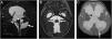
We present the case of a male infant aged 6 months assessed for macrocephaly of 2 month’s duration, born at 38 weeks of gestation and with no history of gestational or perinatal complications. The physical examination revealed that the infant was not able to support his head or roll, milestones that the mother reported he had achieved before the onset of the condition with hypoactivity and vomiting. She also denied any convulsive seizures since birth. Magnetic resonance imaging (MRI) allowed visualization of Blake’s pouch cyst associated with signs of intracranial hypertension (Fig. 1). We referred the patient for placement of a ventriculoperitoneal shunt.

Figure 1.

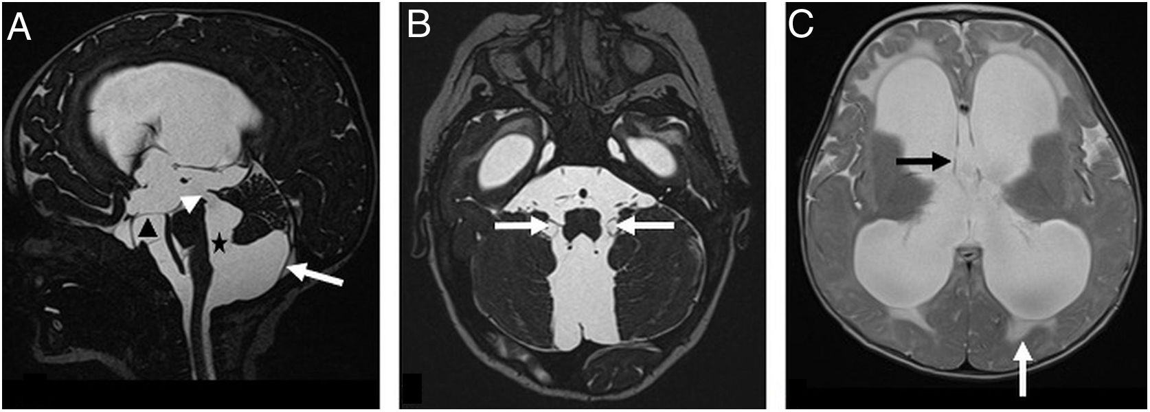
Preoperative MRI in a male infant aged 6 months with severe tetraventricular hydrocephalus. (A) Sagittal image evincing patency of the cerebral aqueduct (white arrowhead) between the enlarged fourth ventricle and Blake’s pouch cyst (black star). Thin posterior cyst wall (white arrow) inferior to the slightly elevated and compressed lobules of the posterior vermis. Concavity of the floor of the third ventricle as a consequence of the cerebrospinal fluid outflow from the ventricles into the cisterns (black arrow). (B) Axial MRI evincing patency of the foramina of Luschka (white arrows). (C) Axial image showing hydrocephalus associated with periventricular interstitial oedema (white arrow) indicative of increased intracranial pressure at the level of the perforated septum pellucidum (black arrow).

(0.11MB).
Financial Disclosure

Authors state that no economic support has been associated with the present study.

Conflicts of Interest

Authors declare no conflict of interest regarding the present study.

References
[1]
L. Mongardi, M. D’Andrea, L. Tosatto, M. Volpin, F. Cultrera.
Endoscopic third ventriculostomy for the treatment of Blake’s pouch cyst in adulthood.
Acta Neurol Belg., 122 (2022), pp. 537-539
[2]
T. Kau, R. Marterer, R. Kottke, R. Birnbacher, J. Gellen, E. Nagy, et al.
Blake's pouch cysts and differential diagnoses in prenatal and postnatal MRI : a pictorial review.
Clin Neuroradiol, 30 (2020), pp. 435-445
[3]
M. Behram, S.C. Oğlak, F. Ölmez, Z. Gedik Özköse, S. Süzen Çaypınar, Y. Başkıran, et al.
Blake’s pouch cyst: prenatal diagnosis and management.
Turk J Obstet Gynecol., 18 (2021), pp. 44-49
Copyright © 2024. Asociación Española de Pediatría
Download PDF
Idiomas
Anales de Pediatría (English Edition)
Article options
Tools