


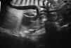
We present the case of a Saharawi pregnant woman with an unremarkable family and personal history. In the current pregnancy, there was evidence of intrauterine growth restriction (<3rd percentile) and fetal blood flow redistribution. The prenatal ultrasound conducted at 38+1 weeks detected an aneurysmal dilatation of the ductus arteriosus (Figs. 1 and 2), so the decision was made to induce labor and admit the infant to the neonatal unit for monitoring due to the potential complications associated with this condition (including spontaneous rupture, embolism, airway erosion, infection, compression of adjacent structures or even death).1,2

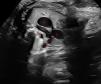
Prenatal ultrasound. 2D mode. Three-vessel and trachea view. The “V” shape produced by the aorta and pulmonary artery appears in the top left of the image. Visualization of the ductus arteriosus with a saccular dilatation with an aneurysmal appearance and a transverse diameter of 10 mm.
Structures: (1) pulmonary artery, (2) aorta, (3) superior vena cava, (4) ductus, (5) trachea.

The postnatal echocardiography confirmed the presence of a ductus arteriosus aneurysm, and the rest of the cardiological evaluation was normal (Appendix A video). During the stay, the infant remained asymptomatic and the aneurysm decreased progressively in size until full closure at day 23 post birth (previous case series have reported favorable outcomes with resolution within 7–35 days). A genetic study was conducted to rule out aortic and vascular diseases, including disorders of connective tissue (such as Marfan or Ehlers-Danlos syndrome) and variants in the ACTA2 gene, which have been associated with aneurysmal dilatation of the great vessels and other systemic malformations.3 We ought to underscore connective tissue disease on account of the increased incidence of complications associated with it.2 In this case, the genetic study was negative. The patient remains asymptomatic to date, with no sonographic abnormalities during the follow-up.
The following is Supplementary data to this article:
Postnatal ultrasound scan video. Modo 2D con doppler color. Aneurysmal dilatation of the ductus arteriosus with opening measuring 4 mm in diameter at the aorta, saccular distension with an ascending thoracic trajectory (maximum diameter, 6 mm) and 2.3 mm in diameter at the pulmonary end. Slow flow at the pulmonary end (maximum velocity, 2.4 m/s).
