array:24 [
  "pii" => "S2341287923001862"
  "issn" => "23412879"
  "doi" => "10.1016/j.anpede.2023.08.004"
  "estado" => "S300"
  "fechaPublicacion" => "2023-11-01"
  "aid" => "3482"
  "copyright" => "Asociación Española de Pediatría"
  "copyrightAnyo" => "2023"
  "documento" => "article"
  "crossmark" => 1
  "subdocumento" => "sco"
  "cita" => "An Pediatr (Barc). 2023;99:366-7"
  "abierto" => array:3 [
    "ES" => true
    "ES2" => true
    "LATM" => true
  ]
  "gratuito" => true
  "lecturas" => array:1 [
    "total" => 0
  ]
  "Traduccion" => array:1 [
    "es" => array:19 [
      "pii" => "S1695403323001510"
      "issn" => "16954033"
      "doi" => "10.1016/j.anpedi.2023.05.020"
      "estado" => "S300"
      "fechaPublicacion" => "2023-11-01"
      "aid" => "3482"
      "copyright" => "Asociación Española de Pediatría"
      "documento" => "article"
      "crossmark" => 1
      "subdocumento" => "sco"
      "cita" => "An Pediatr (Barc). 2023;99:366-7"
      "abierto" => array:3 [
        "ES" => true
        "ES2" => true
        "LATM" => true
      ]
      "gratuito" => true
      "lecturas" => array:1 [
        "total" => 0
      ]
      "es" => array:11 [
        "idiomaDefecto" => true
        "cabecera" => "<span class="elsevierStyleTextfn">IM&#193;GENES EN PEDIATR&#205;A</span>"
        "titulo" => "Una malformaci&#243;n inusual de la caja tor&#225;cica&#58; costilla intrator&#225;cica"
        "tienePdf" => "es"
        "tieneTextoCompleto" => "es"
        "paginas" => array:1 [
          0 => array:2 [
            "paginaInicial" => "366"
            "paginaFinal" => "367"
          ]
        ]
        "titulosAlternativos" => array:1 [
          "en" => array:1 [
            "titulo" => "A rare chest wall deformity&#58; intrathoracic rib"
          ]
        ]
        "contieneTextoCompleto" => array:1 [
          "es" => true
        ]
        "contienePdf" => array:1 [
          "es" => true
        ]
        "resumenGrafico" => array:2 [
          "original" => 0
          "multimedia" => array:7 [
            "identificador" => "fig0010"
            "etiqueta" => "Figura 2"
            "tipo" => "MULTIMEDIAFIGURA"
            "mostrarFloat" => true
            "mostrarDisplay" => false
            "figura" => array:1 [
              0 => array:4 [
                "imagen" => "gr2.jpeg"
                "Alto" => 413
                "Ancho" => 1340
                "Tamanyo" => 85058
              ]
            ]
            "descripcion" => array:1 [
              "es" => "<p id="spar0010" class="elsevierStyleSimplePara elsevierViewall">a&#41; Imagen axial y b&#41; coronal de la TC de t&#243;rax con visualizaci&#243;n de la costilla intrator&#225;cica &#40;puntas de flecha&#41;&#59; c&#41; TC con reconstrucci&#243;n 3 D del pulm&#243;n derecho que muestra la compresi&#243;n del par&#233;nquima pulmonar por la costilla intrator&#225;cica &#40;flechas&#41;&#46;</p> <p id="spar0015" class="elsevierStyleSimplePara elsevierViewall">TC&#58; tomograf&#237;a computarizada&#46;</p>"
            ]
          ]
        ]
        "autores" => array:1 [
          0 => array:2 [
            "autoresLista" => "Furkan Ufuk"
            "autores" => array:1 [
              0 => array:2 [
                "nombre" => "Furkan"
                "apellidos" => "Ufuk"
              ]
            ]
          ]
        ]
      ]
      "idiomaDefecto" => "es"
      "Traduccion" => array:1 [
        "en" => array:9 [
          "pii" => "S2341287923001862"
          "doi" => "10.1016/j.anpede.2023.08.004"
          "estado" => "S300"
          "subdocumento" => ""
          "abierto" => array:3 [
            "ES" => true
            "ES2" => true
            "LATM" => true
          ]
          "gratuito" => true
          "lecturas" => array:1 [
            "total" => 0
          ]
          "idiomaDefecto" => "en"
          "EPUB" => "https://multimedia.elsevier.es/PublicationsMultimediaV1/item/epub/S2341287923001862?idApp=UINPBA00005H"
        ]
      ]
      "EPUB" => "https://multimedia.elsevier.es/PublicationsMultimediaV1/item/epub/S1695403323001510?idApp=UINPBA00005H"
      "url" => "/16954033/0000009900000005/v2_202311111135/S1695403323001510/v2_202311111135/es/main.assets"
    ]
  ]
  "itemSiguiente" => array:19 [
    "pii" => "S2341287923002326"
    "issn" => "23412879"
    "doi" => "10.1016/j.anpede.2023.10.006"
    "estado" => "S300"
    "fechaPublicacion" => "2023-11-01"
    "aid" => "3466"
    "copyright" => "Asociaci&#243;n Espa&#241;ola de Pediatr&#237;a"
    "documento" => "article"
    "crossmark" => 1
    "subdocumento" => "sco"
    "cita" => "An Pediatr &#40;Barc&#41;. 2023;99:368-9"
    "abierto" => array:3 [
      "ES" => true
      "ES2" => true
      "LATM" => true
    ]
    "gratuito" => true
    "lecturas" => array:1 [
      "total" => 0
    ]
    "en" => array:11 [
      "idiomaDefecto" => true
      "cabecera" => "<span class="elsevierStyleTextfn">Images in Paediatrics</span>"
      "titulo" => "Nevoid acanthosis nigricans&#58; a little-known entity"
      "tienePdf" => "en"
      "tieneTextoCompleto" => "en"
      "paginas" => array:1 [
        0 => array:2 [
          "paginaInicial" => "368"
          "paginaFinal" => "369"
        ]
      ]
      "titulosAlternativos" => array:1 [
        "es" => array:1 [
          "titulo" => "Acantosis nigricans nevoide&#58; una entidad poco conocida"
        ]
      ]
      "contieneTextoCompleto" => array:1 [
        "en" => true
      ]
      "contienePdf" => array:1 [
        "en" => true
      ]
      "resumenGrafico" => array:2 [
        "original" => 0
        "multimedia" => array:8 [
          "identificador" => "fig0010"
          "etiqueta" => "Figure 2"
          "tipo" => "MULTIMEDIAFIGURA"
          "mostrarFloat" => true
          "mostrarDisplay" => false
          "figura" => array:1 [
            0 => array:4 [
              "imagen" => "gr2.jpeg"
              "Alto" => 866
              "Ancho" => 3008
              "Tamanyo" => 543026
            ]
          ]
          "detalles" => array:1 [
            0 => array:3 [
              "identificador" => "at0010"
              "detalle" => "Figure "
              "rol" => "short"
            ]
          ]
          "descripcion" => array:1 [
            "en" => "<p id="spar0010" class="elsevierStyleSimplePara elsevierViewall">&#40;A&#41; Oval&#44; light brown plaque with a velvety surface located in the right preauricular region&#46; &#40;B&#41; Dermoscopy allowed visualization of a cerebriform appearance with fissures and crypts &#40;FotoFinder medicam 800 HD digital epiluminescence system&#41;&#46; &#40;C&#41; The histological findings were the same as in case 1 &#40;H-E stain&#44; original magnification &#215;40&#41;&#46;</p>"
          ]
        ]
      ]
      "autores" => array:1 [
        0 => array:2 [
          "autoresLista" => "Sandra Mart&#237;nez-Fern&#225;ndez, Carmen Couselo-Rodr&#237;guez, Diego Soto-Garc&#237;a, Ana Batalla"
          "autores" => array:4 [
            0 => array:2 [
              "nombre" => "Sandra"
              "apellidos" => "Mart&#237;nez-Fern&#225;ndez"
            ]
            1 => array:2 [
              "nombre" => "Carmen"
              "apellidos" => "Couselo-Rodr&#237;guez"
            ]
            2 => array:2 [
              "nombre" => "Diego"
              "apellidos" => "Soto-Garc&#237;a"
            ]
            3 => array:2 [
              "nombre" => "Ana"
              "apellidos" => "Batalla"
            ]
          ]
        ]
      ]
    ]
    "idiomaDefecto" => "en"
    "Traduccion" => array:1 [
      "es" => array:9 [
        "pii" => "S1695403323001273"
        "doi" => "10.1016/j.anpedi.2023.05.014"
        "estado" => "S300"
        "subdocumento" => ""
        "abierto" => array:3 [
          "ES" => true
          "ES2" => true
          "LATM" => true
        ]
        "gratuito" => true
        "lecturas" => array:1 [
          "total" => 0
        ]
        "idiomaDefecto" => "es"
        "EPUB" => "https://multimedia.elsevier.es/PublicationsMultimediaV1/item/epub/S1695403323001273?idApp=UINPBA00005H"
      ]
    ]
    "EPUB" => "https://multimedia.elsevier.es/PublicationsMultimediaV1/item/epub/S2341287923002326?idApp=UINPBA00005H"
    "url" => "/23412879/0000009900000005/v1_202311240521/S2341287923002326/v1_202311240521/en/main.assets"
  ]
  "itemAnterior" => array:19 [
    "pii" => "S2341287923002314"
    "issn" => "23412879"
    "doi" => "10.1016/j.anpede.2023.10.005"
    "estado" => "S300"
    "fechaPublicacion" => "2023-11-01"
    "aid" => "3456"
    "copyright" => "Asociaci&#243;n Espa&#241;ola de Pediatr&#237;a"
    "documento" => "article"
    "crossmark" => 1
    "subdocumento" => "sco"
    "cita" => "An Pediatr &#40;Barc&#41;. 2023;99:364-5"
    "abierto" => array:3 [
      "ES" => true
      "ES2" => true
      "LATM" => true
    ]
    "gratuito" => true
    "lecturas" => array:1 [
      "total" => 0
    ]
    "en" => array:11 [
      "idiomaDefecto" => true
      "cabecera" => "<span class="elsevierStyleTextfn">Images in Paediatrics</span>"
      "titulo" => "Short stature and scoliosis&#58; revealing signs of ultrarare skeletal dysplasia"
      "tienePdf" => "en"
      "tieneTextoCompleto" => "en"
      "paginas" => array:1 [
        0 => array:2 [
          "paginaInicial" => "364"
          "paginaFinal" => "365"
        ]
      ]
      "titulosAlternativos" => array:1 [
        "es" => array:1 [
          "titulo" => "Talla baja y escoliosis&#58; datos reveladores de displasia esquel&#233;tica ultrarara"
        ]
      ]
      "contieneTextoCompleto" => array:1 [
        "en" => true
      ]
      "contienePdf" => array:1 [
        "en" => true
      ]
      "resumenGrafico" => array:2 [
        "original" => 0
        "multimedia" => array:8 [
          "identificador" => "fig0005"
          "etiqueta" => "Figure 1"
          "tipo" => "MULTIMEDIAFIGURA"
          "mostrarFloat" => true
          "mostrarDisplay" => false
          "figura" => array:1 [
            0 => array:4 [
              "imagen" => "gr1.jpeg"
              "Alto" => 1222
              "Ancho" => 755
              "Tamanyo" => 56405
            ]
          ]
          "detalles" => array:1 [
            0 => array:3 [
              "identificador" => "at0005"
              "detalle" => "Figure "
              "rol" => "short"
            ]
          ]
          "descripcion" => array:1 [
            "en" => "<p id="spar0005" class="elsevierStyleSimplePara elsevierViewall">Complete X-ray of the spine &#40;7<span class="elsevierStyleHsp" style=""></span>years and 7 months&#41;&#58; multiple vertebral and rib malformations&#46; Scoliosis with high dorsal curve and mild convexity to the right&#46;</p>"
          ]
        ]
      ]
      "autores" => array:1 [
        0 => array:2 [
          "autoresLista" => "Silvia Rodr&#237;guez del Rosario, Silvia Modamio-H&#248;ybj&#248;r, Karen E&#46; Heath, M&#46; Pilar Bahillo-Curieses"
          "autores" => array:4 [
            0 => array:2 [
              "nombre" => "Silvia"
              "apellidos" => "Rodr&#237;guez del Rosario"
            ]
            1 => array:2 [
              "nombre" => "Silvia"
              "apellidos" => "Modamio-H&#248;ybj&#248;r"
            ]
            2 => array:2 [
              "nombre" => "Karen E&#46;"
              "apellidos" => "Heath"
            ]
            3 => array:2 [
              "nombre" => "M&#46; Pilar"
              "apellidos" => "Bahillo-Curieses"
            ]
          ]
        ]
      ]
    ]
    "idiomaDefecto" => "en"
    "Traduccion" => array:1 [
      "es" => array:9 [
        "pii" => "S1695403323001170"
        "doi" => "10.1016/j.anpedi.2023.05.004"
        "estado" => "S300"
        "subdocumento" => ""
        "abierto" => array:3 [
          "ES" => true
          "ES2" => true
          "LATM" => true
        ]
        "gratuito" => true
        "lecturas" => array:1 [
          "total" => 0
        ]
        "idiomaDefecto" => "es"
        "EPUB" => "https://multimedia.elsevier.es/PublicationsMultimediaV1/item/epub/S1695403323001170?idApp=UINPBA00005H"
      ]
    ]
    "EPUB" => "https://multimedia.elsevier.es/PublicationsMultimediaV1/item/epub/S2341287923002314?idApp=UINPBA00005H"
    "url" => "/23412879/0000009900000005/v1_202311240521/S2341287923002314/v1_202311240521/en/main.assets"
  ]
  "en" => array:14 [
    "idiomaDefecto" => true
    "cabecera" => "<span class="elsevierStyleTextfn">Images in Paediatrics</span>"
    "titulo" => "A rare chest wall deformity&#58; Intrathoracic rib"
    "tieneTextoCompleto" => true
    "paginas" => array:1 [
      0 => array:2 [
        "paginaInicial" => "366"
        "paginaFinal" => "367"
      ]
    ]
    "autores" => array:1 [
      0 => array:3 [
        "autoresLista" => "Furkan Ufuk"
        "autores" => array:1 [
          0 => array:3 [
            "nombre" => "Furkan"
            "apellidos" => "Ufuk"
            "email" => array:1 [
              0 => "furkan.ufuk@hotmail.com"
            ]
          ]
        ]
        "afiliaciones" => array:1 [
          0 => array:2 [
            "entidad" => "Department of Radiology&#44; Pamukkale University&#44; Denizli&#44; T&#252;rkiye"
            "identificador" => "aff0005"
          ]
        ]
      ]
    ]
    "titulosAlternativos" => array:1 [
      "es" => array:1 [
        "titulo" => "Una malformaci&#243;n inusual de la caja tor&#225;cica&#58; costilla intrator&#225;cica"
      ]
    ]
    "resumenGrafico" => array:2 [
      "original" => 0
      "multimedia" => array:8 [
        "identificador" => "fig0005"
        "etiqueta" => "Figure 1"
        "tipo" => "MULTIMEDIAFIGURA"
        "mostrarFloat" => true
        "mostrarDisplay" => false
        "figura" => array:1 [
          0 => array:4 [
            "imagen" => "gr1.jpeg"
            "Alto" => 665
            "Ancho" => 755
            "Tamanyo" => 77514
          ]
        ]
        "detalles" => array:1 [
          0 => array:3 [
            "identificador" => "at0005"
            "detalle" => "Figure "
            "rol" => "short"
          ]
        ]
        "descripcion" => array:1 [
          "en" => "<p id="spar0005" class="elsevierStyleSimplePara elsevierViewall">Anterior&#8211;posterior chest radiograph showing an abnormally oriented rib in the right side &#40;arrowheads&#41;&#46;</p>"
        ]
      ]
    ]
    "textoCompleto" => "<span class="elsevierStyleSections"><p id="par0005" class="elsevierStylePara elsevierViewall">A girl aged 5 years presented with a cough and difficulty breathing of one week&#8217;s duration&#46; She had no known disease&#44; and the physical examination was unremarkable&#46; The posterior-anterior chest radiograph showed an abnormally oriented rib in the upper right side of the chest &#40;<a class="elsevierStyleCrossRef" href="#fig0005">Fig&#46; 1</a>&#41;&#46; The patient underwent a low-dose CT scan of the chest without contrast for further evaluation&#46; The CT scan revealed the presence of a bifid intrathoracic rib protruding into the lung parenchyma &#40;<a class="elsevierStyleCrossRef" href="#fig0010">Fig&#46; 2</a>&#44; <a class="elsevierStyleCrossRef" href="#sec0005">Movie</a>&#41;&#46; Since the patient&#8217;s symptoms were mild&#44; conservative treatment was initiated and a follow-up visit scheduled&#46;</p><elsevierMultimedia ident="fig0005"></elsevierMultimedia><elsevierMultimedia ident="fig0010"></elsevierMultimedia><p id="par0010" class="elsevierStylePara elsevierViewall">An intrathoracic rib&#44; also known as a thoracic rib or slipping rib syndrome&#44; is an extremely rare anomaly of the chest wall in which a normal&#44; supernumerary or bifid rib is located within the chest cavity&#46;<a class="elsevierStyleCrossRef" href="#bib0005"><span class="elsevierStyleSup">1</span></a> The clinical manifestations of intrathoracic rib can include sharp or dull pain&#44; difficulty breathing or shortness of breath and a palpable lump or bulge near the involved rib&#46;<a class="elsevierStyleCrossRef" href="#bib0005"><span class="elsevierStyleSup">1</span></a> The management of this type of malformation typically involves a combination of conservative measures and&#44; in severe cases&#44; surgical intervention&#46;<a class="elsevierStyleCrossRef" href="#bib0010"><span class="elsevierStyleSup">2</span></a> A chest CT scan allows precise visualization of the tubular shape and curvature of the intrathoracic rib and can be used to guide surgery&#46;<a class="elsevierStyleCrossRefs" href="#bib0005"><span class="elsevierStyleSup">1&#44;2</span></a></p></span>"
    "pdfFichero" => "main.pdf"
    "tienePdf" => true
    "apendice" => array:1 [
      0 => array:1 [
        "seccion" => array:1 [
          0 => array:4 [
            "apendice" => "<p id="par0020" class="elsevierStylePara elsevierViewall">The following are Supplementary data to this article&#58;<elsevierMultimedia ident="upi0005"></elsevierMultimedia></p>"
            "etiqueta" => "Appendix A"
            "titulo" => "Supplementary data"
            "identificador" => "sec0010"
          ]
        ]
      ]
    ]
    "multimedia" => array:3 [
      0 => array:8 [
        "identificador" => "fig0005"
        "etiqueta" => "Figure 1"
        "tipo" => "MULTIMEDIAFIGURA"
        "mostrarFloat" => true
        "mostrarDisplay" => false
        "figura" => array:1 [
          0 => array:4 [
            "imagen" => "gr1.jpeg"
            "Alto" => 665
            "Ancho" => 755
            "Tamanyo" => 77514
          ]
        ]
        "detalles" => array:1 [
          0 => array:3 [
            "identificador" => "at0005"
            "detalle" => "Figure "
            "rol" => "short"
          ]
        ]
        "descripcion" => array:1 [
          "en" => "<p id="spar0005" class="elsevierStyleSimplePara elsevierViewall">Anterior&#8211;posterior chest radiograph showing an abnormally oriented rib in the right side &#40;arrowheads&#41;&#46;</p>"
        ]
      ]
      1 => array:8 [
        "identificador" => "fig0010"
        "etiqueta" => "Figure 2"
        "tipo" => "MULTIMEDIAFIGURA"
        "mostrarFloat" => true
        "mostrarDisplay" => false
        "figura" => array:1 [
          0 => array:4 [
            "imagen" => "gr2.jpeg"
            "Alto" => 413
            "Ancho" => 1340
            "Tamanyo" => 85136
          ]
        ]
        "detalles" => array:1 [
          0 => array:3 [
            "identificador" => "at0010"
            "detalle" => "Figure "
            "rol" => "short"
          ]
        ]
        "descripcion" => array:1 [
          "en" => "<p id="spar0010" class="elsevierStyleSimplePara elsevierViewall">&#40;a&#41; Axial and &#40;b&#41; coronal chest CT images evincing the intrathoracic rib &#40;arrowheads&#41;&#46; &#40;c&#41; Three-dimensional volume reconstruction chest CT image of the right lung demonstrates the parenchymal indentation of the intrathoracic rib &#40;arrows&#41;&#46;</p>"
        ]
      ]
      2 => array:7 [
        "identificador" => "upi0005"
        "etiqueta" => "Movie 1"
        "tipo" => "MULTIMEDIAECOMPONENTE"
        "mostrarFloat" => false
        "mostrarDisplay" => true
        "Ecomponente" => array:3 [
          "fichero" => "mmc1.mp4"
          "ficheroTamanyo" => 547985
          "Video" => array:2 [
            "mp4" => array:5 [
              "fichero" => "mmc1.m4v"
              "poster" => "mmc1.jpg"
              "tiempo" => 0
              "alto" => 0
              "ancho" => 0
            ]
            "flv" => array:5 [
              "fichero" => "mmc1.flv"
              "poster" => "mmc1.jpg"
              "tiempo" => 0
              "alto" => 0
              "ancho" => 0
            ]
          ]
        ]
        "descripcion" => array:1 [
          "en" => "<p id="spar0015" class="elsevierStyleSimplePara elsevierViewall">Three-dimensional reconstruction CT scan of the chest showing the protrusion of the intrathoracic rib into the chest cavity&#46;</p>"
        ]
      ]
    ]
    "bibliografia" => array:2 [
      "titulo" => "References"
      "seccion" => array:1 [
        0 => array:2 [
          "identificador" => "bibs0005"
          "bibliografiaReferencia" => array:2 [
            0 => array:3 [
              "identificador" => "bib0005"
              "etiqueta" => "1"
              "referencia" => array:1 [
                0 => array:2 [
                  "contribucion" => array:1 [
                    0 => array:2 [
                      "titulo" => "Intrathoracic rib&#58; rare rib anomaly&#44; review of the literature and proposal for classification"
                      "autores" => array:1 [
                        0 => array:2 [
                          "etal" => false
                          "autores" => array:4 [
                            0 => "X&#46; Xue"
                            1 => "S&#46; Zhao"
                            2 => "K&#46; Li"
                            3 => "B&#46; Zhao"
                          ]
                        ]
                      ]
                    ]
                  ]
                  "host" => array:1 [
                    0 => array:2 [
                      "doi" => "10.7150/ijms.63828"
                      "Revista" => array:6 [
                        "tituloSerie" => "Int J Med Sci"
                        "fecha" => "2021"
                        "volumen" => "18"
                        "paginaInicial" => "3800"
                        "paginaFinal" => "3807"
                        "link" => array:1 [
                          0 => array:2 [
                            "url" => "https://www.ncbi.nlm.nih.gov/pubmed/34790056"
                            "web" => "Medline"
                          ]
                        ]
                      ]
                    ]
                  ]
                ]
              ]
            ]
            1 => array:3 [
              "identificador" => "bib0010"
              "etiqueta" => "2"
              "referencia" => array:1 [
                0 => array:2 [
                  "contribucion" => array:1 [
                    0 => array:2 [
                      "titulo" => "Single-port thoracoscopic intrathoracic rib resection&#58; a case description"
                      "autores" => array:1 [
                        0 => array:2 [
                          "etal" => false
                          "autores" => array:5 [
                            0 => "C&#46; Wang"
                            1 => "G&#46; Hu"
                            2 => "B&#46; Min"
                            3 => "Z&#46; Cheng"
                            4 => "X&#46; Zuo"
                          ]
                        ]
                      ]
                    ]
                  ]
                  "host" => array:1 [
                    0 => array:2 [
                      "doi" => "10.21037/qims-22-421"
                      "Revista" => array:6 [
                        "tituloSerie" => "Quant Imaging Med Surg"
                        "fecha" => "2023"
                        "volumen" => "13"
                        "paginaInicial" => "507"
                        "paginaFinal" => "511"
                        "link" => array:1 [
                          0 => array:2 [
                            "url" => "https://www.ncbi.nlm.nih.gov/pubmed/36620144"
                            "web" => "Medline"
                          ]
                        ]
                      ]
                    ]
                  ]
                ]
              ]
            ]
          ]
        ]
      ]
    ]
  ]
  "idiomaDefecto" => "en"
  "url" => "/23412879/0000009900000005/v1_202311240521/S2341287923001862/v1_202311240521/en/main.assets"
  "Apartado" => array:4 [
    "identificador" => "77701"
    "tipo" => "SECCION"
    "en" => array:2 [
      "titulo" => "Images in Paediatrics"
      "idiomaDefecto" => true
    ]
    "idiomaDefecto" => "en"
  ]
  "PDF" => "https://static.elsevier.es/multimedia/23412879/0000009900000005/v1_202311240521/S2341287923001862/v1_202311240521/en/main.pdf?idApp=UINPBA00005H&text.app=https://analesdepediatria.org/"
  "EPUB" => "https://multimedia.elsevier.es/PublicationsMultimediaV1/item/epub/S2341287923001862?idApp=UINPBA00005H"
]
Share
Journal Information
Vol. 99. Issue 5.
Pages 366-367 (1 November 2023)
Vol. 99. Issue 5.
Pages 366-367 (1 November 2023)
Images in Paediatrics
Full text access
A rare chest wall deformity: Intrathoracic rib
Una malformación inusual de la caja torácica: costilla intratorácica
Visits
1658
Furkan Ufuk
Department of Radiology, Pamukkale University, Denizli, Türkiye
This item has received
Article information
Full Text
Bibliography
Download PDF
Statistics
Figures (2)
Additional material (1)
Full Text

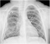
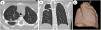
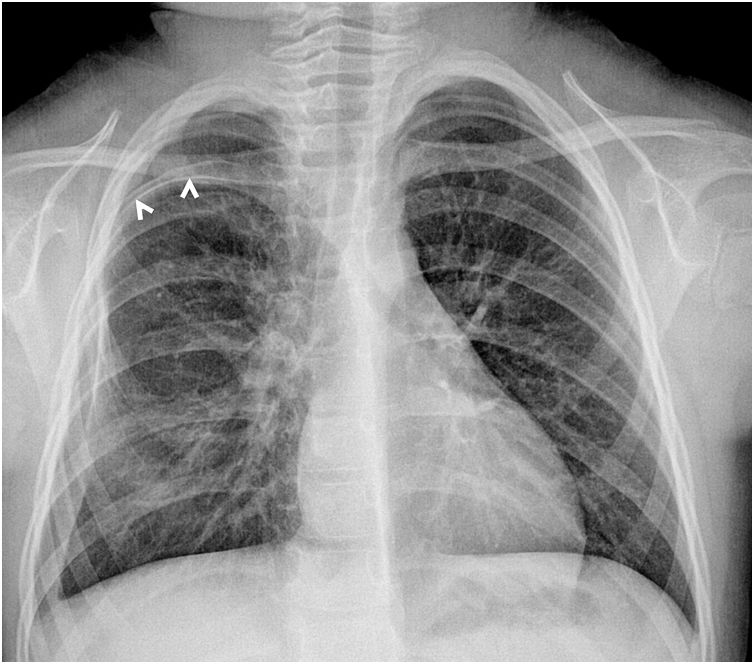
A girl aged 5 years presented with a cough and difficulty breathing of one week’s duration. She had no known disease, and the physical examination was unremarkable. The posterior-anterior chest radiograph showed an abnormally oriented rib in the upper right side of the chest (Fig. 1). The patient underwent a low-dose CT scan of the chest without contrast for further evaluation. The CT scan revealed the presence of a bifid intrathoracic rib protruding into the lung parenchyma (Fig. 2, Movie). Since the patient’s symptoms were mild, conservative treatment was initiated and a follow-up visit scheduled.

Figure 1.

Anterior–posterior chest radiograph showing an abnormally oriented rib in the right side (arrowheads).

(0.07MB).
Figure 2.

(a) Axial and (b) coronal chest CT images evincing the intrathoracic rib (arrowheads). (c) Three-dimensional volume reconstruction chest CT image of the right lung demonstrates the parenchymal indentation of the intrathoracic rib (arrows).

(0.08MB).

An intrathoracic rib, also known as a thoracic rib or slipping rib syndrome, is an extremely rare anomaly of the chest wall in which a normal, supernumerary or bifid rib is located within the chest cavity.1 The clinical manifestations of intrathoracic rib can include sharp or dull pain, difficulty breathing or shortness of breath and a palpable lump or bulge near the involved rib.1 The management of this type of malformation typically involves a combination of conservative measures and, in severe cases, surgical intervention.2 A chest CT scan allows precise visualization of the tubular shape and curvature of the intrathoracic rib and can be used to guide surgery.1,2

Appendix A
Supplementary data

The following are Supplementary data to this article:

(0.52MB)

Three-dimensional reconstruction CT scan of the chest showing the protrusion of the intrathoracic rib into the chest cavity.

References
[1]
X. Xue, S. Zhao, K. Li, B. Zhao.
Intrathoracic rib: rare rib anomaly, review of the literature and proposal for classification.
Int J Med Sci, 18 (2021), pp. 3800-3807
[2]
C. Wang, G. Hu, B. Min, Z. Cheng, X. Zuo.
Single-port thoracoscopic intrathoracic rib resection: a case description.
Quant Imaging Med Surg, 13 (2023), pp. 507-511
Copyright © 2023. Asociación Española de Pediatría
Download PDF
Idiomas
Anales de Pediatría (English Edition)
Article options
Tools
Supplemental materials