
Limited dorsal myeloschisis is a primary neurulation defect resulting from incomplete disjunction of the cutaneous and neural ectoderm. It manifests with a cutaneous lesion and a fibroneural stalk linking the lesion to the underlying spinal cord.1,2
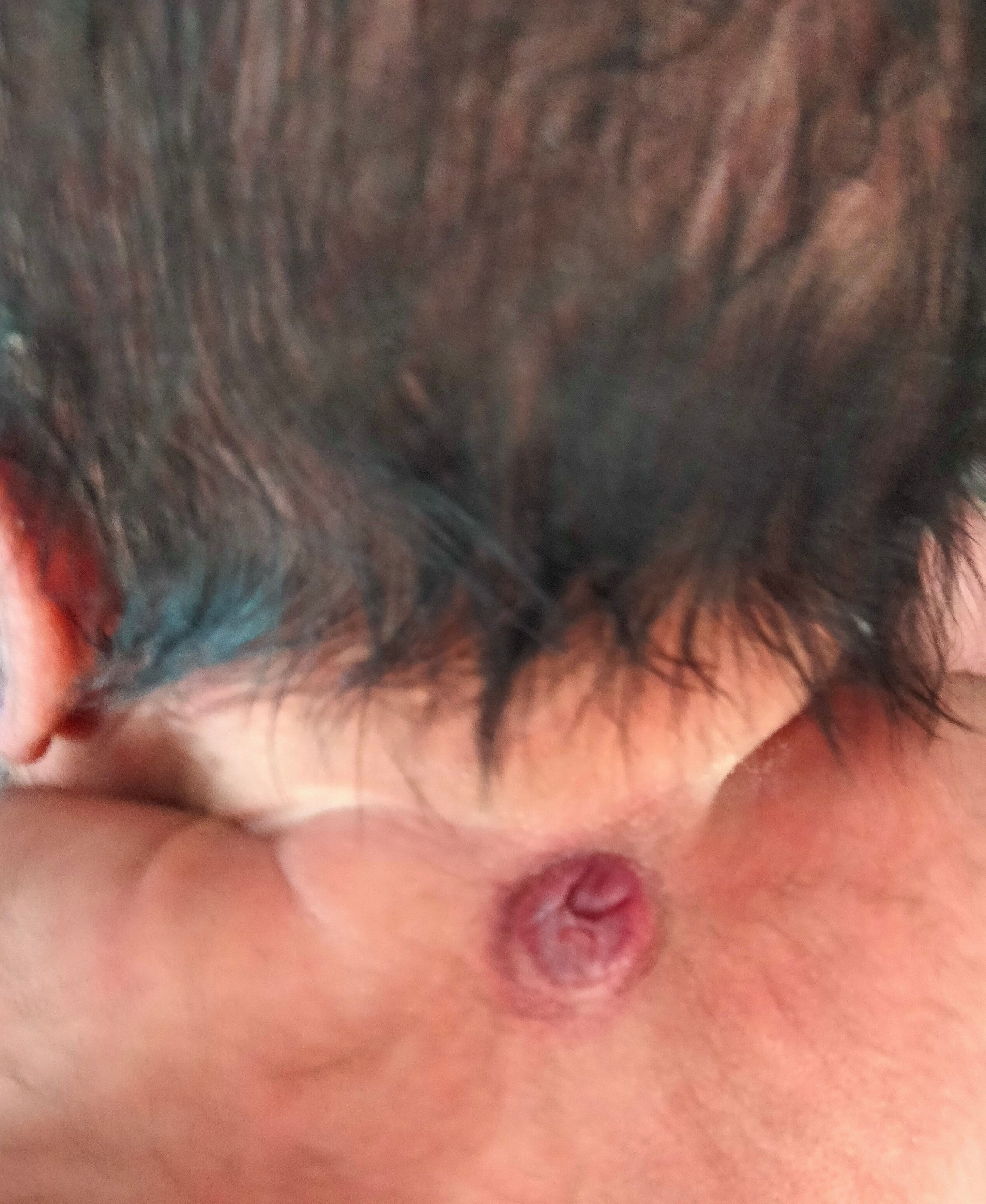
We present the case of a male neonate born with a circular lesion on the neck that measured 2&#¿;×&#¿;3&#¿;cm and was covered by a flaccid membrane (Fig. 1). The prenatal ultrasound findings had been normal. Treatment with folic acid was interrupted in the early months of pregnancy.

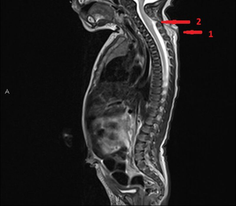
The next day, the lesion had enlarged and contained serosanguineous fluid, increasing in size in association with increasing intraabdominal pressure (Fig. 2). A cervical ultrasound scan revealed a hypoechoic tract that extended to a small area with absence of fusion of the posterior laminae. An MRI scan allowed visualization of an ascending tract that reached the thecal sac, with mild dilation at the level of C4-C5, with slight tethering of the spinal cord toward it including the meninges (Fig. 3). At 4 days post birth, the patient underwent dissection and closure of the defect. The histological examination of the specimen revealed the presence of glioneuronal tissue in the mid and deep dermis positive for S100 protein and glial fibrillary acidic protein (GFAP), in addition to a fibrous tract with cells positive for GFAP.


