Journal Information
Vol. 100. Issue 1.
Pages 77-78 (01 January 2024)
Vol. 100. Issue 1.
Pages 77-78 (01 January 2024)
Images in Paediatrics
Full text access
Annular grouped blisters on an infant
Lesiones anulares ampollosas en lactante
Visits
409
Jana Codina Barbosaa,
Corresponding author
jana.codina@sjd.es

Corresponding author.
, Ares Benavent Puiggenéa, Mariana Álvarez Vukovb, Eulàlia Baselga Torresc, Yolanda Fernández Santervásd
a Servicio de Pediatría, Hospital Sant Joan de Déu, Universidad de Barcelona, Barcelona, Spain
b Servicio de Anatomía Patológica, Hospital Sant Joan de Déu, Universidad de Barcelona, Barcelona, Spain
c Servicio de Dermatología Pediátrica, Hospital Sant Joan de Déu, Universidad de Barcelona, Barcelona, Spain
d Área de Urgencias, Hospital Sant Joan de Déu Barcelona, Esplugues de Llobregat, Barcelona, Spain
This item has received
Article information
Full Text
Bibliography
Download PDF
Statistics
Figures (3)
Show moreShow less
Full Text

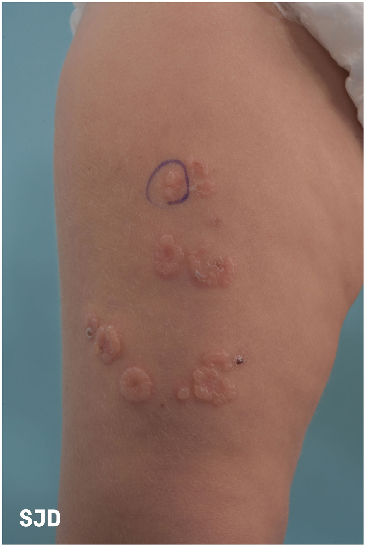
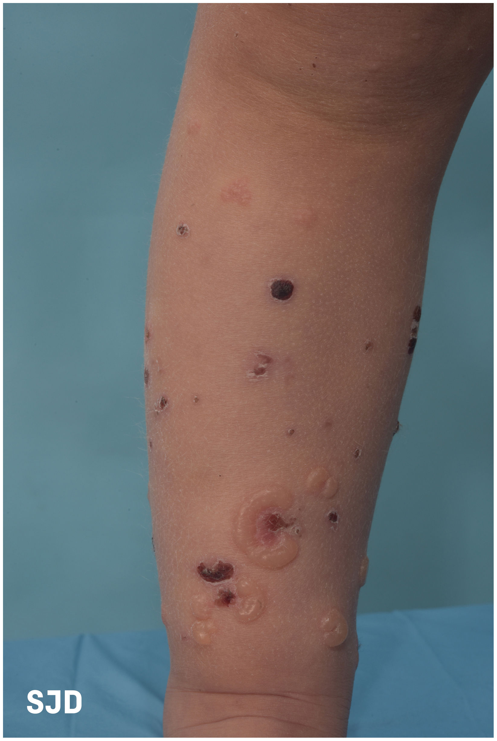
A patient aged 20 months with no relevant medical history was brought to the emergency department for assessment of skin lesions with onset 15 days prior. The examination (Figs. 1 and 2) revealed the presence of tense vesicles and blisters clustered around macules or crusts with an arrangement resembling a rosette. The patient was afebrile and there was no mucosal involvement. No exposure to pharmaceuticals or recent vaccination reported.

Figure 1.

Bulging blisters in a rosette arrangement.

(0.15MB).
Figure 2.

Formation of crusts in some lesions.

(0.28MB).

Linear immunoglobulin A bullous dermatosis was suspected. The findings of the histological examination and direct immunofluorescence (DIF) confirmed the diagnosis: subepidermal bullae and visualization on DIF of continuous linear deposition of IgA at the dermoepidermal junction (Fig. 3).

Figure 3.

Direct immunofluorescence for detection of immunoglobulin A: continuous the dermoepidermal junction.

(0.14MB).

Treatment was initiated with prednisone, adding dapsone after 15 days at a dose of 1mg/kg/day with tapering off of prednisone until its discontinuation. The levels of glucose-6-phosphate dehydrogenase remained in the normal range, and the patient had a favourable outcome.

Linear immunoglobulin A bullous is an acquired blistering dermatosis of an autoimmune aetiology,1 although there can be cases induced by drugs. The main diseases that need to be considered in the differential diagnosis are dermatitis herpetiformis, bullous pemphigoid, lichen planus and toxic epidermal necrolysis.1–3 It is important for paediatricians to know how to recognise the typical rosette or ‘cluster of jewels’ pattern, although the pathognomonic feature is the linear deposit of IgA along the basal membrane. Dapsone is the treatment of choice, and the course of disease is characterised by exacerbation and remission periods until its full resolution.1

Funding

This research did not receive any external funding.

References
[1]
G. Fortuna, M.P. Marinkovich.
Linear immunoglobulin A bullous dermatosis.
[2]
V. Fuentelsaz del Barrio, M. Campos Domínguez.
Dermatosis IgA lineal de la infancia.
Rev Pediatr Aten Primaria, 15 (2013), pp. 141-145
[3]
P. Joly, M. Castel.
Dermatosis IgA linear.
Copyright © 2023. Asociación Española de Pediatría
Idiomas
Anales de Pediatría (English Edition)
Article options
Tools
es en

¿Es usted profesional sanitario apto para prescribir o dispensar medicamentos?

Are you a health professional able to prescribe or dispense drugs?