

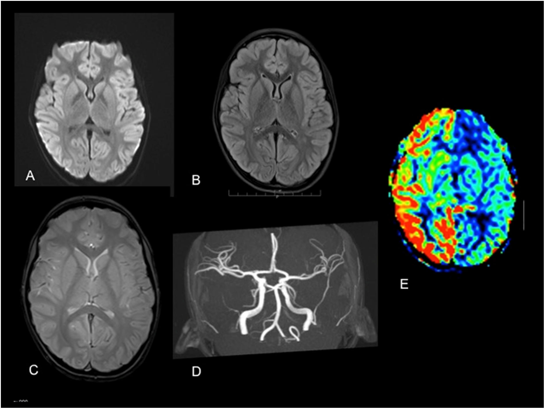
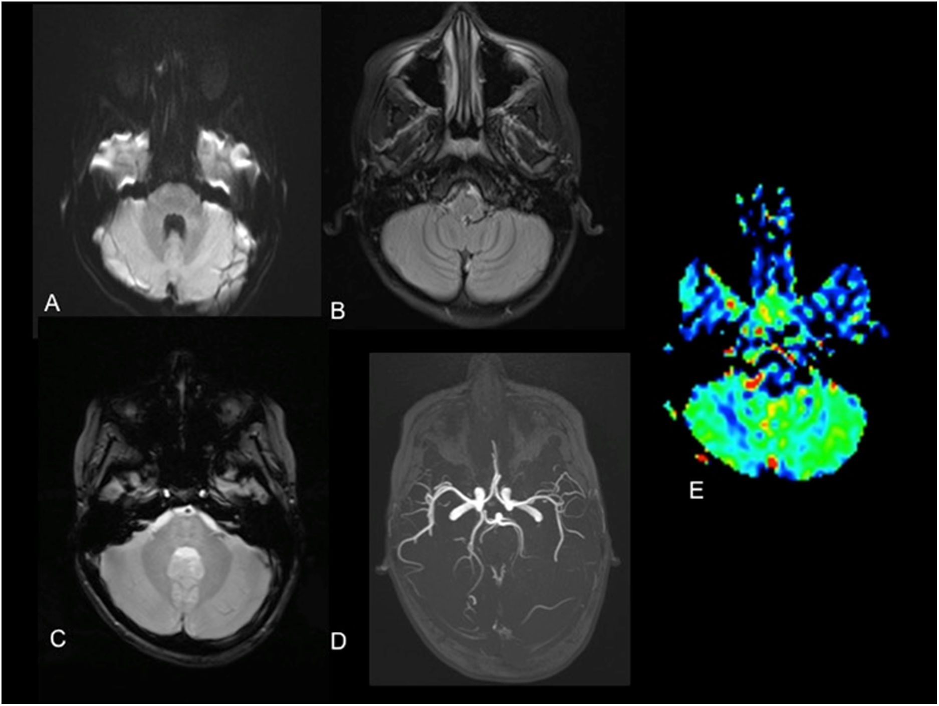
A right-handed girl aged 7 years presented to our hospital with an episode of generalised muscular rigidity with jerking movements in the right upper extremity, disconnection syndrome and relaxation of sphincters. The patient received intravenous benzodiazepines in the emergency department, which achieved resolution of the seizures. Since the patient continued to have a low Glasgow Comma Scale score, aphasia and immobilization of the extremity, the stroke code was activated. A perfusion magnetic resonance imaging scan performed 2 h after onset showed decreased blood flow in the left hemisphere (Fig. 1) and the right cerebellar hemisphere (Fig. 2), with preserved perfusion in the basal ganglia. The electroencephalogram evinced an abundance of epileptiform discharges in the right-side anterior frontotemporal region. Since Todd paralysis was suspected, an intravenous bolus of levetiracetam was administered to the patient, which achieved full resolution of the symptoms in 24 h.
 diffusion sequence, B) FLAIR sequence, C) gradient echo sequence and D) TOF angiography revealing no abnormalities. E) Decreased blood flow in left hemisphere with preservation of left basal ganglia in ASL perfusion sequence. ASL, arterial spin labelling: FLAIR, fluid-attenuated inversion recovery; TOF, time of flight.")
Magnetic resonance imaging at the level of the basal ganglia. A) diffusion sequence, B) FLAIR sequence, C) gradient echo sequence and D) TOF angiography revealing no abnormalities. E) Decreased blood flow in left hemisphere with preservation of left basal ganglia in ASL perfusion sequence.
ASL, arterial spin labelling: FLAIR, fluid-attenuated inversion recovery; TOF, time of flight.
 diffusion sequence, B) FLAIR sequence, C) gradient echo sequence and D) TOF angiography revealing no abnormalities. E) Decreased blood flow in right cerebellar hemisphere in ASL perfusion sequence. ASL, arterial spin labelling: FLAIR, fluid-attenuated inversion recovery; TOF, time of flight.")
Magnetic resonance imaging at the level of the cerebellar hemispheres. A) diffusion sequence, B) FLAIR sequence, C) gradient echo sequence and D) TOF angiography revealing no abnormalities. E) Decreased blood flow in right cerebellar hemisphere in ASL perfusion sequence.
ASL, arterial spin labelling: FLAIR, fluid-attenuated inversion recovery; TOF, time of flight.
Todd paralysis, also known as post-ictal paralysis, is a condition of unknown aetiology and variable duration1 manifesting with focal weakness after a seizure,2 although, if it involves the dominant side, it can manifest with aphasia in addition to motor impairment,3 as was the case of our patient, and the main differential diagnosis is stroke. Perfusion imaging can evince a reduction in blood flow that does not match a vascular territory, sparing the basal ganglia, and crossed cerebellar diaschisis due to disconnection secondary to the transient metabolic changes in the contralateral corticopontocerebellar pathway.

