We present the new diagnostic techniques for use in primary care pediatrics, which have been included in the Spanish public health system nationwide portfolio of services, pursuant to the Royal Decree published in the Official State Gazette (BOE) on May 13, 2025. Specifically, the newly included techniques are tympanometry, transcutaneous bilirubin measurement, rapid testing for respiratory viruses, measurement of C-reactive protein in capillary blood, rapid testing for allergies, instrument-based vision screening, dermoscopy, and point-of-care ultrasound.
Presentamos las nuevas técnicas diagnósticas de aplicación en Pediatría de Atención primaria, que se han introducido en la nueva cartera de servicios común del Sistema Nacional de Salud, según Real decreto que recoge la orden ministerial publicada en el BOE de 13-5-2025. Se detallan específicamente: timpanometría, bilirrubinometría transcutánea, test de diagnóstico rápido para virus respiratorios, determinación de proteina C reactiva en sangre capilar, test rápido para patología alérgica, refractometría, dermatoscopia y ecografía clínica.
The capacity of the primary care (PC) system to resolve presenting problems without referral to higher levels of care needs to improve, and this requires the allocation of additional resources to pediatricians to allow them, in specific circumstances, to use diagnostic tests the results of which can modify epidemiological, diagnostic and therapeutic decisions, thus contributing to a more rational use of the resources of the public health care system and improving its efficiency.1
On May 13, 2025, the Royal Decree by which the Ministry of Health mandates the reform of the nationwide PC services portfolio of the National Health System (NHS) was published in the Boletín Oficial del Estado (BOE, Official State Gazette).2 This portfolio was developed by consensus by a working group appointed by the Ministry in 2022 and composed of representatives of Spanish PC scientific societies (in the field of pediatrics: the AEPAP, represented by César García Vera, and the SEPEAP, represented by the TECDIAP [Working Group on Diagnostic Technologies in Primary Care]), as well as the PC directorates of each of the autonomous communities.
The most relevant techniques newly included in the services portfolio are the following:
- -
Tympanometry.
- -
Transcutaneous bilirubin measurement.
- -
Rapid diagnostic test (RDT) for respiratory viruses, including influenza, respiratory syncytial virus (RSV), adenovirus, SARS-CoV-2, and Epstein-Barr virus.
- -
Capillary C-reactive protein (CRP) test.
- -
Allergy testing with measurement of specific immunoglobulin E (IgE).
- -
Refractometry.
- -
Dermoscopy.
- -
Point-of-care ultrasound (POCUS).
This article does not cover tests that were already available in previous versions of the portfolio, such as the rapid strep test (included since 2018).
Below, we proceed to describe these techniques in the order they were mentioned, discussing their potential impact on pediatric PC practice.
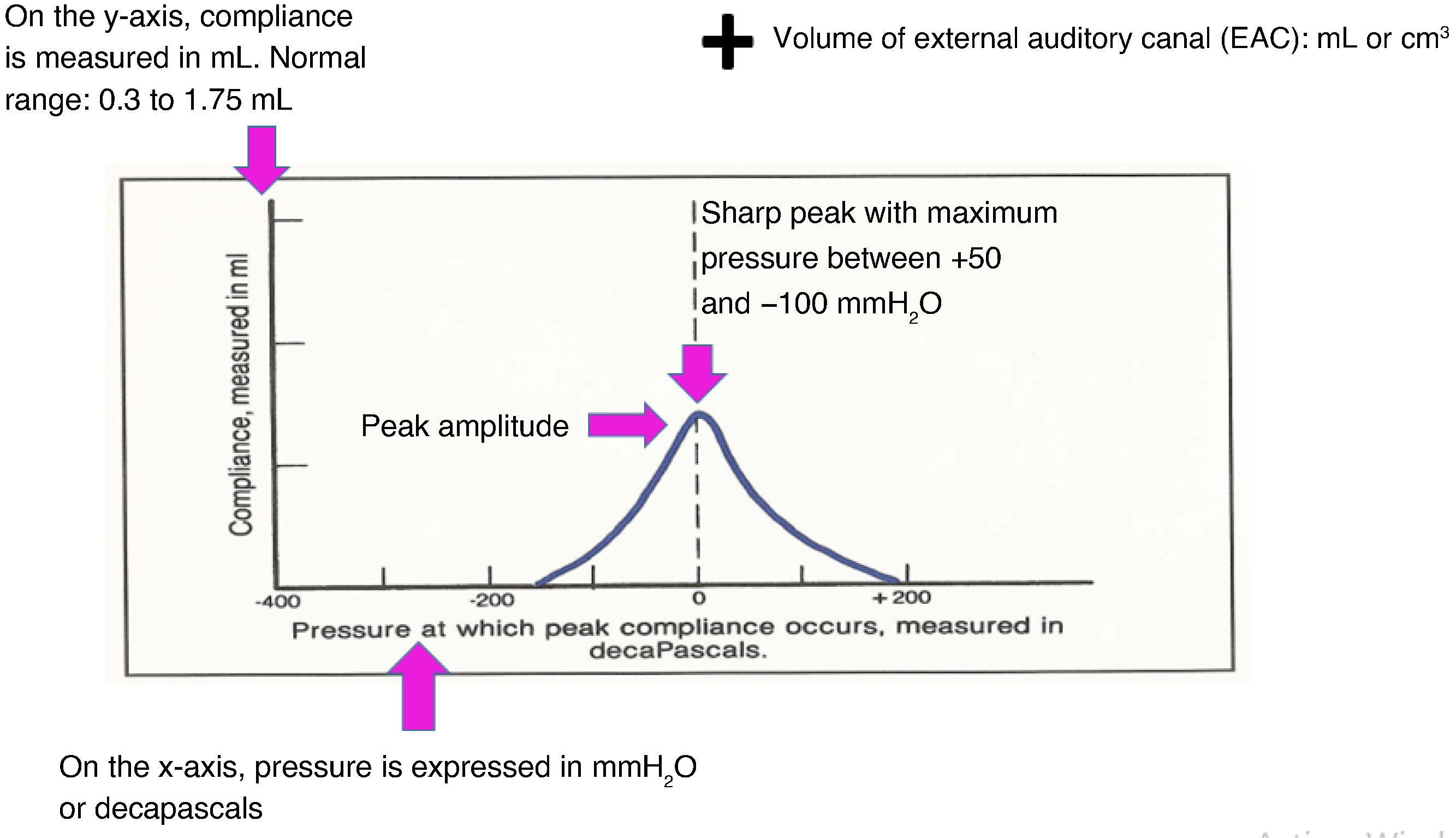
TympanometryIt is a noninvasive technique with substantial validation3,4 that offers a sensitivity and specificity similar to those of tympanocentesis for confirmation of the presence of exudate in the middle ear. The output of the technique is a curve call tympanogram (Fig. 1).

It has a specificity of 82% to 89% and a sensitivity of 95% to 100%.
Tympanometry allows:
- •
Detection of fluid in the middle ear.
- •
Assessment of eustachian tube function.
- •
Measurement of the volume of the external auditory canal (EAC) to assess for perforation and the functioning of ear drainage tubes.
In practice, clinicians must know how to interpret 3 curves:
- -
Normal: centered and with a sharp tent-like peak. It rules out the presence of exudate.
- -
Flat or undetectable curve: allows diagnosis of suppurative otitis media or otitis media with effusion, or, in the presence of compatible symptoms, acute otitis media (AOM).
- -
Peak shifted to the left: negative pressure in the middle ear. It is indicative of eustachian tube dysfunction or ototubaritis.
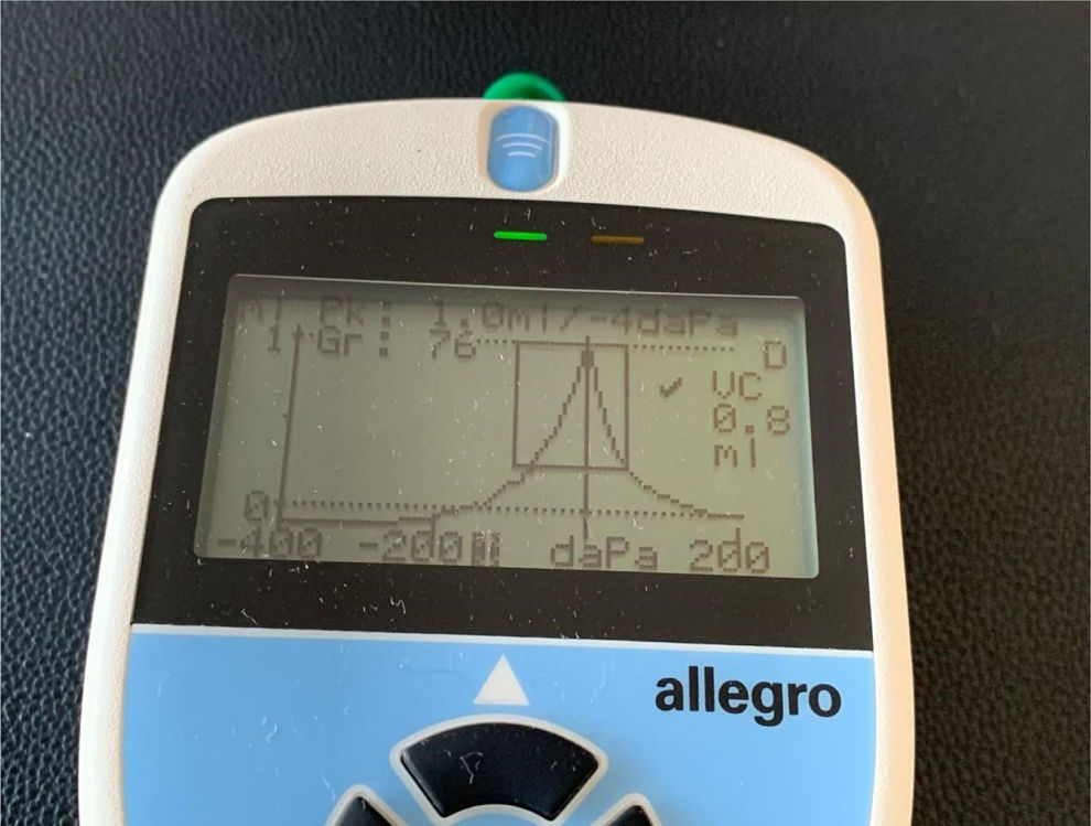
Tympanometry alone cannot be used to diagnose AOM, which requires the presence of signs and symptoms of acute inflammation. The technique can also be used to check whether the eardrum is perforated and to verify that transtympanic drains are functioning properly. Both handheld and desktop tympanometers (Fig. 2) are currently available.

In conclusion, tympanometry is an essential diagnostic technique, given the difficulties in diagnosing otitis media by means of conventional otoscopy and the need to make the diagnosis as objective as possible due to its implications for treatment and follow-up.5–7
Transcutaneous bilirubin measurementThe risks posed by toxic bilirubin levels and the poor correlation between visual appearance and actual serum levels, especially in infants born before 38 weeks,8 warrants the development and implementation of guidelines regarding bilirubin measurement, monitoring, and management with phototherapy,9,10 and some authors have even proposed universal screening before discharge,11,12 so it would be very useful for PC providers to have the means to objectively measure bilirubin.
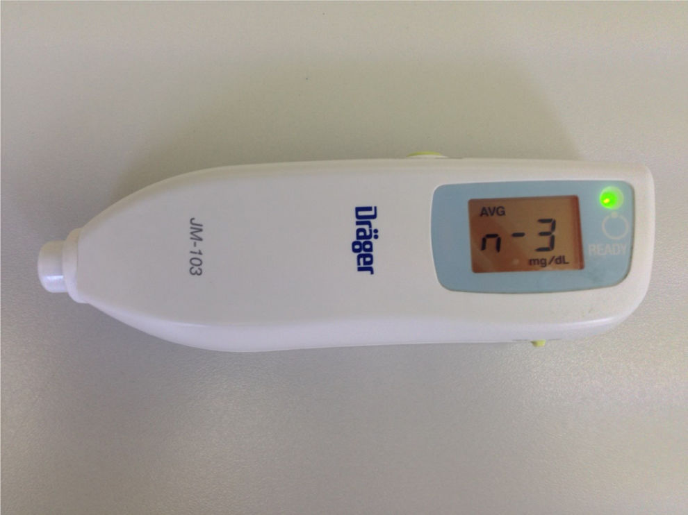
There is evidence of a strong correlation between transcutaneous bilirubin (TcB) (Fig. 3) and serum bilirubin values, making TcB measurement a useful screening method at the PC level for identifying which infants should be referred to a hospital for performance of a serum bilirubin test to determine whether phototherapy is indicated.13–15 The technique has been validated for use in newborn infants weighing more than 1000 g. In preterm infants with lower birth weights, the margin of discrepancy between TcB and serum bilirubin values (± 3 to 4 mg/dL) is considered too large to recommend this technique to guide decision-making.

In the PC setting, TcB measurement is recommended for all newborns with jaundice. A baseline test and a follow-up test at 24 h should be performed. The patient should be referred to the hospital in the following cases:
- •
TcB > 12 mg/dL in term infants.
- •
TcB > 15 mg/dL in preterm infants.
- •
Increase in TcB > 5 mg/dL in 24 h.
It is recommended that each PC team consult the nomogram of the corresponding referral hospital in order to establish common referral criteria.
In summary, we believe that the transcutaneous bilirubinometer is an essential tool for pediatric PC teams.
Rapid diagnostic test (RDT) for respiratory virusesIn some seasons, the hospitalization rate due to influenza in children aged less than 2 years is comparable to that of people aged 65 years and older.16 Differentiating the flu from other viral respiratory infections based solely on the clinical presentation is challenging in children, with a level of difficulty that is inversely associated with age. The ability to make an etiological diagnosis could reduce the frequency of unscheduled return visits associated with nonspecific febrile illnesses, and thus the risk that a viral infection might result in performance of low-yield diagnostic tests, referral to the local emergency department, or unnecessary antibiotic treatment—risks that increase in direct proportion to the days of illness.
A landmark study in Spain17 analyzed the impact of rapid influenza diagnostic tests (RIDTs) in the population aged less than 6 years. It concluded that their use achieved a reduction in both return visits and antibiotic treatment, in agreement with international studies on the subject.
Immunochromatographic RIDTs are convenient, easy to use, inexpensive, and offer read times of 5 to 15 minutes. A metaanalysis18 of 159 studies in children and adults comparing RIDT and culture, 35% of which were conducted during the 2009 pandemic, found an average sensitivity of 62.3% and an average specificity of 98%. The sensitivity was higher in children (66.6%) and for influenza A (64.6%).
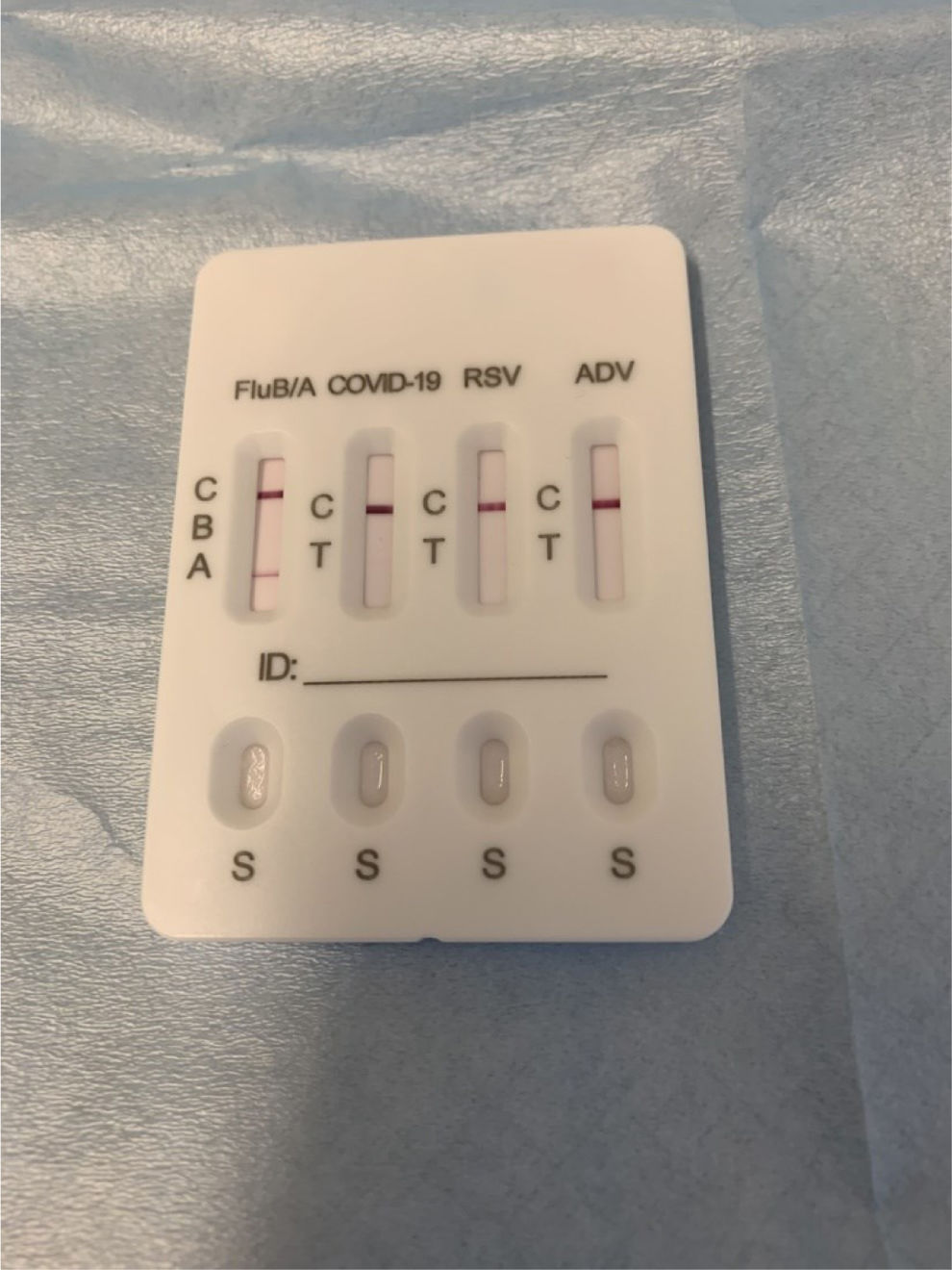
The optimal window to perform a RIDT is between 12 and 48 h from the onset of symptoms, and it should always be done in the first 4–5 days. Before 12 h, it can lead to false negatives due to a low antigen load. Different RIDTs can detect influenza A alone, influenza B alone, or both in the same cassette. The test included in the updated portfolio is a multiplex cassette for detection of influenza as well as RSV, SARS-CoV-2 and adenovirus antigen in a single sample (Fig. 4).
Other RDTs for respiratory infections: RSV, adenovirus, SARS-CoV-2, and infectious mononucleosisRapid diagnostic test for respiratory syncytial virus (RSV)
Although clinical practice guidelines do not recommend the routine performance of RDTs for detection of RSV, our experience with its use has evinced the following benefits in the management of bronchiolitis19:
- •
Facilitating the necessary isolation in the common situation of mild bronchiolitis in a young child managed at home, during which the child should not go to the nursery or preschool, a recommendation that is frequently not given when the child is afebrile and in good general health.
- •
In most cases, the use of bronchodilators does not modify the course of RSV-related bronchiolitis, but some patients show clear improvement with bronchodilators during the therapeutic trial, in which case their prescription for at-home treatment is indicated; on the other hand, the diagnosis of bronchiolitis caused by RSV should be considered a criterion to withhold the use of corticosteroids at the PC level.
- •
Rapid diagnostic testing can be useful in the conservative management of febrile infants with bronchiolitis. The presence of RSV is associated with a low probability of severe bacterial infection, and its documentation allows deferral of other diagnostic tests in the initial assessment.
- •
The availability of this test at the PC level reduces the use of antibiotics in the outpatient management of pneumonia. In cases of clinically suspected community-acquired pneumonia in a child aged less than 2 years during the RSV season, a positive RDT should be determinant in the decision to initially withhold antibiotherapy.
Having this test available at the PC level would be useful in the following situations20:
- -
In the differential diagnosis of suspected streptococcal pharyngitis with a negative rapid strep test versus infectious mononucleosis.
- -
For the etiological diagnosis of viral pneumonia: adenovirus is the third leading cause following RSV and parainfluenza.
- -
To reduce the use of antibiotics in pneumonia.
The RDT for Epstein-Barr virus B is based on the detection of IgM heterophile antibodies through immunochromatography. The sample of capillary blood can be easily obtained through a finger prick. Studies comparing enzyme immunoassay and hemagglutination tests have found a sensitivity and specificity greater than 99%.21 New rapid techniques measure IgG against capsid viral antigen (CVA), which also appears early in the infection, and are more sensitive compared to the Paul-Bunnell heterophile antibody test.
Capillary C-reactive protein (CRP) testC-reactive protein, with a cut-off value of 20 mg/L, offers a diagnostic accuracy similar to that of procalcitonin (PCT).22,23 It has a window period of approximately 8 h that constitutes a limitation in the evaluation of very early febrile episodes, which are commonly encountered in everyday clinical practice.
Over the past 20 years, the following thresholds have been established to assess the probable etiology of the infectious process 24:
- •
20 mg/L: probably viral.
- •
30−70 mg/L: uncertain etiology. In some viral infections, CRP can be elevated to these levels.
- •
>70 mg/L: probably bacterial.
C-reactive protein thresholds are only indicative cannot be used in isolation to guide decision-making without considering the clinical presentation. This test has been proposed as a useful indicator in the etiologic differentiation of community-acquired pneumonia, the classification of urinary tract infections (UTIs) as probable upper UTI (> 70 mg/L, acute pyelonephritis) or lower UTI (< 70 mg/L, cystitis), and, above all, the evaluation of fever without source in children at risk of occult bacteremia (OB), and its use is associated with a decrease in unnecessary referrals to hospital, optimization of management at the PC level, and avoidance of unnecessary tests and, in many cases, of empiric antibiotherapy.
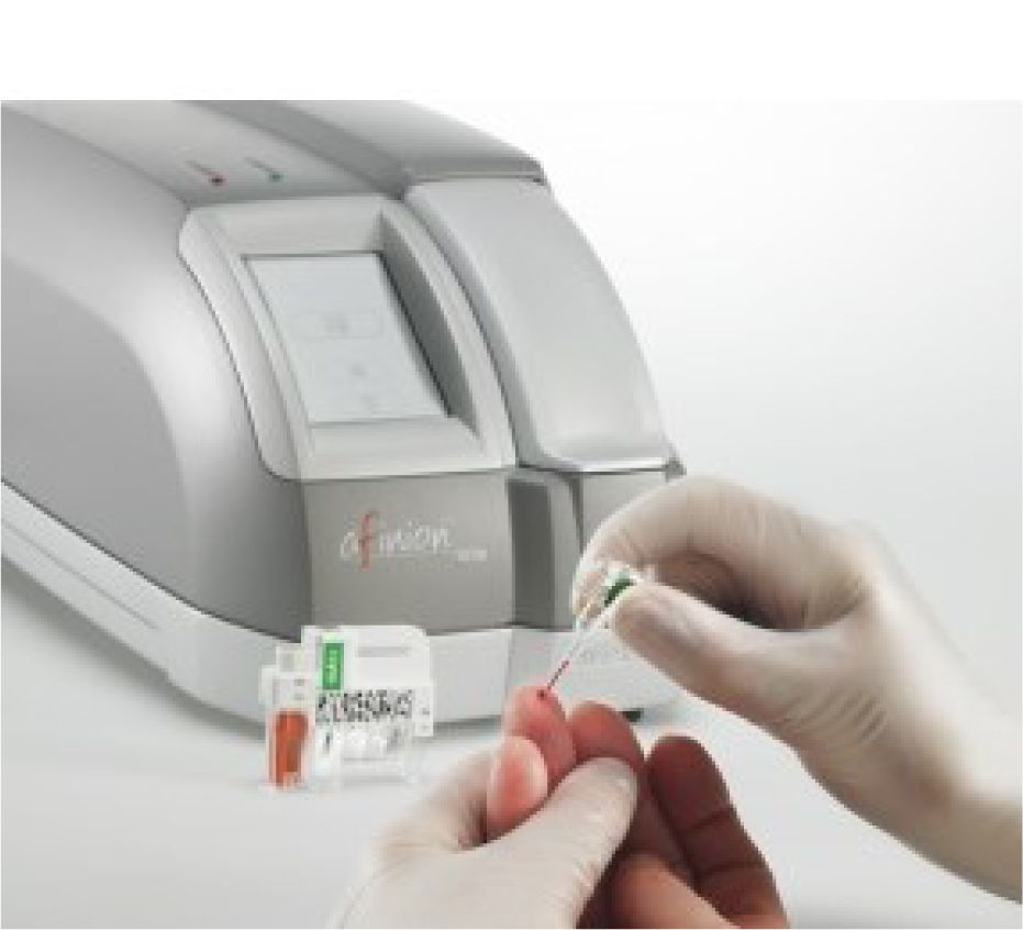
Capillary CRP testing is very convenient for children, inexpensive, and it offers results in just a few minutes, allowing for practical decisions to be made within the same visit. It requires a point-of-care analyzer (Fig. 5).
Rapid diagnostic testing for allergic conditions
In PC practice, rapid allergy tests are useful for identifying patients with allergies, providing guidance on avoidance strategies, and referring patients to a specialist for further evaluation. They can also help rule out sensitivities that improve with age in preschool-aged children with recurrent wheezing.
The rapid allergy test is an in vitro immunoassay for the detection of allergen-specific IgE antibodies in blood, using a panel of 10 inhalants and food allergens from a single capillary blood sample. Its main advantages are its simplicity of use and the short turnaround time (20−30 min).
A multicenter study25 conducted in children aged 1–14 years that compared this rapid test with allergen-specific IgE testing of venous blood found that the kit was appropriate in 96.7% of patients with suspected atopy and its use was associated with a 78.7% decrease in referrals.
It should be considered a first-line test for the evaluation of children with recurrent wheezing at the PC level.
Instrument-based vision screeningMost cases of amblyopia develop between the ages of 1 and 3 years in the context of uncorrected anisometropia or strabismus.
With the aim of detecting these conditions earlier than eye charts allow, new tools have been developed, such as binocular autorefractors, which enable the objective assessment of visual refraction from age 6 months.26
These devices allow screening of children at much younger ages than 3–4 years, and they can detect the most common conditions that increase the risk of amblyopia.
- •
Anisometropia (unequal refractive error in each eye).
- •
Strabismus.
- •
Bilateral refractive errors: congenital myopia, astigmatism, or high hyperopia.
- •
Conditions associated with opacities or leukocoria: cataracts, childhood glaucoma, persistent fetal vasculature, retinoblastoma.
In addition, autorefractors can detect unilateral or bilateral refractive errors (hyperopia, astigmatism, myopia) that, while not associated with the risk of amblyopia, reduce visual acuity in young children, with an impact on learning.
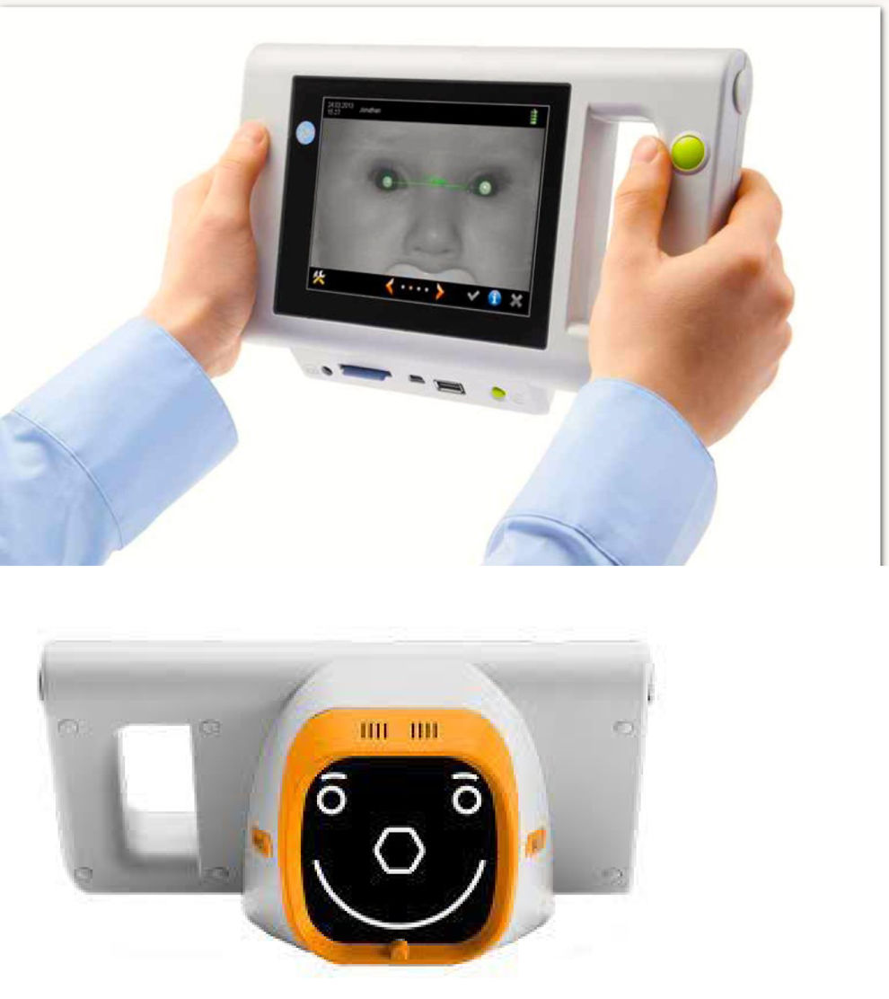
Autorefractors (Fig. 6) are instruments that automatically and objectively measure the refraction of both eyes simultaneously in patients aged 6 months and older. They perform automated infrared binocular photoretinoscopy and require very little patient cooperation.

The mean sensitivity is 81%–92%, with the advantage that screening can be performed as early as 6 months of age, and it takes a fraction of the time compared to testing visual acuity with eye charts.
In Spain,27 one multicenter prospective study was carried out in four PC centers in Barcelona in patients aged 18–30 months. In this study, the sensitivity for detecting risk factors for amblyopia was 89%, and the specificity was 91%. Approximately 50% of patients referred between ages 18 and 30 months required optical correction following their initial evaluation by the ophthalmology team, and 40% of those with a confirmed refractive error for which correction was not initially indicated were scheduled for follow-up ophthalmological visits.
In conclusion, autorefractors are the instrument of choice for screening for refractive errors in children aged 1–4 years. Furthermore, they enable screening of all children with developmental and communication disorders who may not always receive appropriate care because they are “uncooperative”.
Dermoscopy in primary care pediatricsDermoscopy is an in vivo, noninvasive diagnostic technique that has revolutionized the management of pediatric skin diseases.28 Its integration into the PC service portfolio marks a paradigm shift toward “precision pediatrics,” serving as a crucial link between clinical practice and anatomical pathology by eliminating the phenomena of light refraction and reflection on the skin.29 Because it is a quick and painless technique, it is ideal for pediatric patients, as it causes neither psychological distress nor physical discomfort.
The fundamental advantage of dermoscopy is that it allows visualization of structures beyond the stratum corneum of the skin, including deeper layers of the epidermis, the dermo-epidermal junction, and the papillary dermis, which are invisible to the naked eye.30 In pediatric patients, this technique significantly increases the diagnostic sensitivity of PC providers, even with minimal training. The use of simplified algorithms, such as Soyer’s 3-point checklist, is recommended to facilitate its implementation in primary care.
The implementation of pediatric dermoscopy is based on three core principles:
- 1
Minimization of iatrogenesis: It improves secondary prevention of childhood melanoma by avoiding unnecessary biopsies and surgical excisions, thereby reducing the psychological impact, pain, and scarring in children.
- 2
Optimization of patient flow: The technique has a negative predictive value of nearly 100% in confirming the benign nature of a lesion, reducing family anxiety and minimizing unnecessary referrals to dermatology specialists.
- 3
Clinical versatility: Its usefulness extends beyond pigmented lesions, facilitating the diagnosis and monitoring of a wide range of conditions, such as parasitic infections,31 inflammatory lesions, alopecia (trichoscopy),32 and nail disorders (onychoscopy).
It is a cost-effective technique with a manageable learning curve.
Pediatric point-of-care ultrasoundPoint-of-care ultrasound (POCUS), also known as bedside ultrasound, has established itself as an invaluable tool in pediatric clinical practice. Considered an extension of the physical examination, it allows for the integration of real-time imaging, facilitating answers to specific diagnostic questions and improving decision-making within the same visit.33 Its inclusion in the portfolio of services offered by the National Health System provides key institutional support for its deployment.
Lung ultrasound stands out for its high sensitivity in the diagnosis of pneumonia and other respiratory infections, avoiding radiation exposure and enabling ongoing monitoring. In addition, it improves diagnostic accuracy and helps optimize treatment, reducing the use of antibiotics and other diagnostic tests.34,35
Abdominal ultrasound can be used in the initial evaluation of numerous clinical conditions, such as acute abdomen, intussusception, hypertrophic pyloric stenosis, or renal and urinary tract disorders, facilitating immediate decision-making regarding management or the need for specialized care.36
In musculoskeletal assessments, ultrasound offers a high diagnostic yield and is useful for detecting fractures, joint effusions, synovitis, or fluid collections.37 In addition, it helps guide the initial diagnosis toward specific traumatological or rheumatological conditions (vascular or inflammatory, in the presence of compatible manifestations) based on indirect findings, streamlining clinical decision-making and the ordering of additional tests. It is also the technique of choice for screening for developmental hip dysplasia in infants, which consolidates its role in the early detection of orthopedic disorders in children.
Brain and cranial ultrasound, especially in infants, allows the initial assessment of intracranial and extracranial conditions with a good diagnostic yield in certain situations.38,39 Its use in PC facilitates the detection of structural abnormalities and the monitoring of the progression of various lesions, avoiding more complex or invasive tests and contributing to the early detection of significant neurological conditions, even in cases with nonspecific clinical manifestations.
The neck ultrasound is also a useful tool in the investigation of common neck conditions; it is particularly helpful for assessment of lymphadenopathy and thyroid disorders and in the initial evaluation of cervical masses, providing rapid guidance for clinical management.40
Cutaneous ultrasound allows for the visualization of the skin and subcutaneous tissue. It is useful for characterizing superficial lesions, cysts, foreign bodies, inflammatory or infectious conditions, and vascular malformations, providing information on their depth, extent, and vascularization that increases the accuracy of the initial diagnosis at the PC clinic.
The integration of POCUS in PC improves the diagnostic and therapeutic process by enabling clinically significant findings and guiding decision-making at the point of care. This results in a reduction in unnecessary referrals, a more rational use of health care resources, and greater efficiency of the health care system. It also shortens diagnostic turnaround times and increases satisfaction among patients and health care professionals by providing immediate answers in the PC clinic—a particularly valuable aspect in a care setting focused on accessibility and problem-solving capacity.
The use of POCUS in pediatric care in Spain is still limited and unequal. Although many providers have access to ultrasound equipment, its actual use is lower than expected, primarily due to a lack of specific training and heavy workloads. However, there is broad consensus on the need to incorporate ultrasound into pediatric training, reflecting a growing interest in the technique and its future potential.
Training is a key factor for ensuring the proper use of POCUS. Structured programs including theoretical instruction, supervised practical training, and competency assessment need to be developed. The use of ultrasound by pediatricians with specific training on the technique is acceptable provided that adequate training, appropriate application in the clinical setting, and quality control mechanisms are in place to ensure patient safety.
In short, pediatric POCUS is an accessible, safe, and highly valuable tool that enhances the resolution capacity of primary care. Its inclusion in the portfolio of services and the available evidence support its integration into clinical practice. Its implementation requires adequate equipment, structured training, and allocation of sufficient time for its performance. Its implementation will contribute to our progress towards a more effective, efficient, and patient-centered pediatric care practice and the alignment of our health care model with the Sustainable Development Goals. Furthermore, its adoption offers a strategic opportunity to strengthen the role of PC within the health care system.
ConclusionThe gradual implementation of the techniques introduced in this update to the nationwide portfolio of pediatric PC services within the National Health System will mark a decisive shift in pediatric practice in this care setting, leading to a significant increase in the rate of resolution at the PC level and the satisfaction of health care professionals.
The authors have no conflicts of interest to declare.