

Aplasia cutis congenita (ACC) is a rare and heterogenous disorder, with an estimated rate of incidence 1–3 to 10,000 births.1 It is characterized by the absence of a skin portion and sometimes subcutaneous tissue and even bone.1–3 Usually ACC presents itself as a solitary scalp defect but it can also manifest as multiple scalp lesions or it can affect any other part of the body.1,2 In some situations, ACC can be associated with other physical anomalies or malformation syndromes.2
We present five cases of ACC of the scalp that occurred in a very short period of time (four months) with no apparent relation between them. In all the cases, the parents were less than 35 years old, with no history of maternal infections or drug intake during pregnancy.
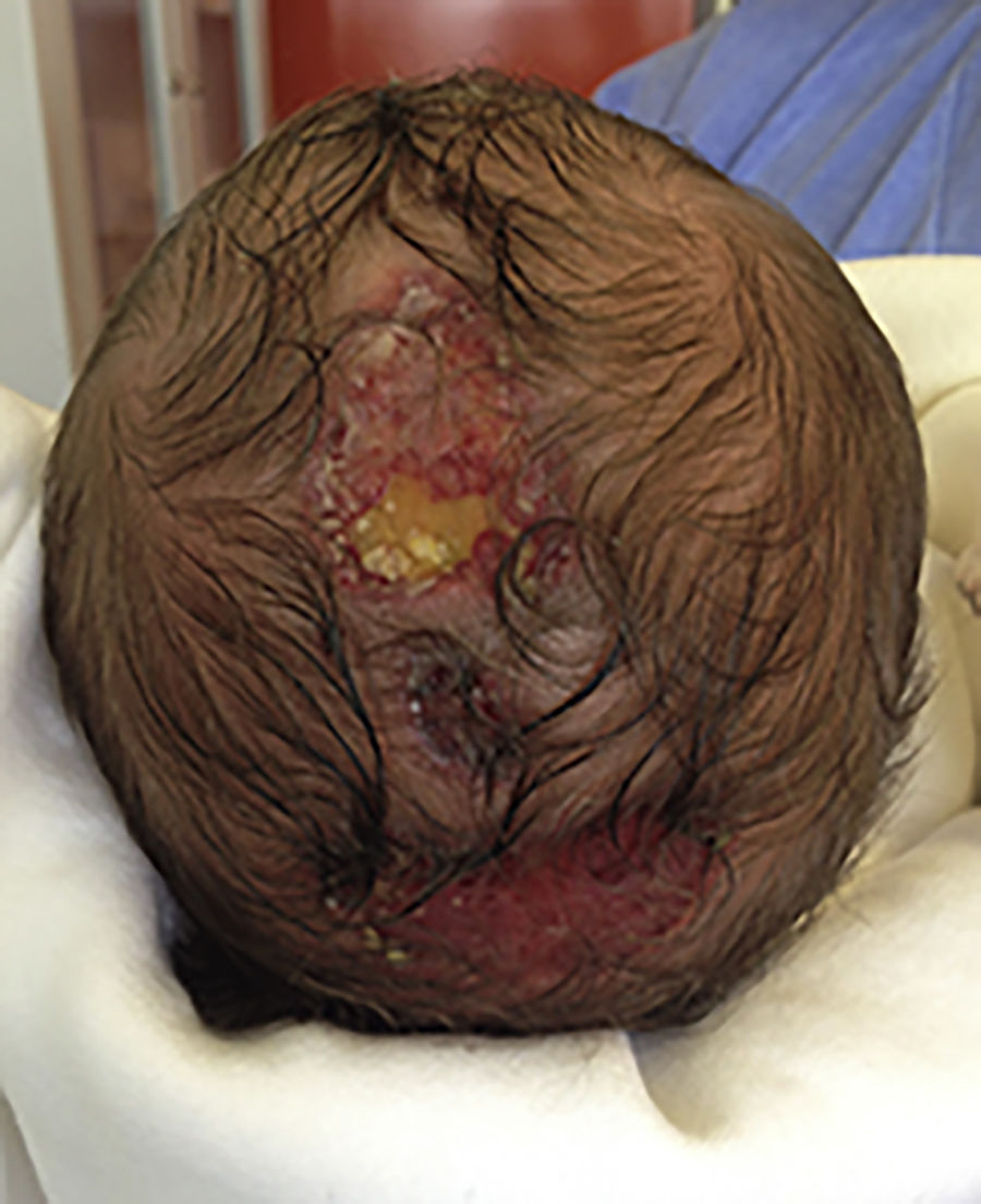
The first one was a male neonate from an uneventful pregnancy with normal obstetric ultrasounds (US). The infant was born at term by spontaneous vaginal delivery and, immediately after birth, he was noted with three oval lesions in the median region of the scalp, the biggest measuring 4cm×3cm, all with well-defined margins (Fig. 1). The cranial US did not show any central nervous system haemorrhage, neither signs of infection or sinus thrombosis and cranial computer tomography revealed bone integrity of the scalp.

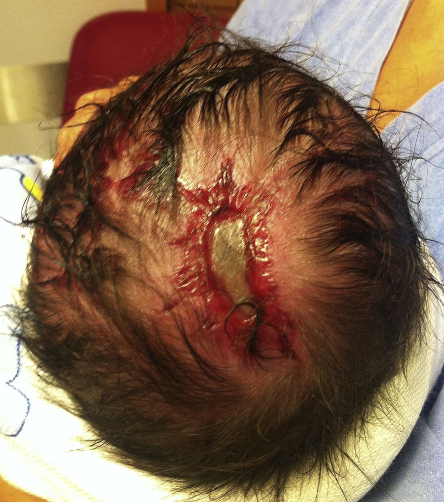
The second was also a male term neonate, but in this case, there was a history of dichorionic diamniotic twin pregnancy with spontaneous foetal death of one foetus at 15 weeks gestational age (papyraceous foetus). The surviving twin was born with a single oval shaped scalp lesion with 3cm×2cm (Fig. 2). The cranial US was also normal and a cranial magnetic resonance confirmed the absence of central nervous system defect, which are more prevalent in the surviving neonate twin of a papyraceous foetus.

The last three neonates, one male and two females, were born with a small circular defect on the scalp vertex, between 3 and 5mm. The male neonate's gestation was complicated by gestational diabetes and at 35 weeks an emergency caesarean section was precipitated by an umbilical cord prolapse, bringing about an extremely difficult extraction; additionally, his mother exhibited a small alopecia lesion since birth. Female neonates were term gestations, with no maternal comorbidities and no abnormal events during pregnancy, and were born from spontaneous vaginal deliveries. Because of the small size of the lesions, these three only performed cranial US, that did not reveal any alterations.
In all five cases, the treatment was conservative. Twice daily, sterile swabs soaked in a combined biguanide polyhexanide and betaine surfactant solution were used to clean the lesions, followed by a fusidic acid ointment until clinical improvement. There were no acute complications registered and healing occurred in all the cases in 6–12 weeks, with an alopecia in place of the previous skin defect.
In most of cases, ACC is an isolated lesion but it can also be a part of various malformation syndromes.1 Any part of the body can be affected, but the scalp, usually the vertex, is the more frequent location of the defect occurring in 70–90% of cases, with underlying bone defects in 20–30%.1
The pathogenic mechanism is still unclear, but the condition has been suggested to occur as a result of disrupted development or degeneration of skin in utero.4 Several studies describe an association with foetus papyraceous, but others attribute the defect to genetics, placental infarcts, teratogenic substances, intrauterine infections, trauma, vascular compromise, amniogenesis, adhesions of the amniotic membrane to the foetal skin, among other theories.4 In the presented cases, we just found possible mechanisms in two of them (papyraceous foetus in one and a possible genetic/traumatic labour in another).
The treatment is not consensual. The conservative approach is the first choice in most cases, but some authors defend that the surgical approach can be a better option when the defect is larger than 4cm or when there is bone absence.1 Otherwise, some studies demonstrate that even larger lesions can be successfully treated with conservative treatment,3,5 as seen in our first case.
Local infection or bleeding are the main acute complications, but infections of the central nervous system and thrombosis or haemorrhages of the sagittal sinus are the most serious complications and they can be fatal. These are related with the size of the lesion and the involvement of the bone.2 Alopecia and hypertrophic scars are the more frequent late complications, usually not reversible.2 In the presented cases, there were no acute complications but alopecia occurred in all of them.
Please cite this article as: Almeida S, Rodrigues F, Coelho S, Bicho MA. Cinco casos de aplasia cutis congénita. An Pediatr (Barc). 2018;89:212–213.
Previous presentation: Presented at the 7th Aveiro-Viseu Pediatrics Conference, June 16 and 17, 2016, Viseu, Portugal.
- Documento de manejo clínico del paciente pediátrico con infección por SARS-CoV-2.
(Actualizado el 26 de Noviembre de 2020) - Test de diagnóstico rápido en las consultas de Pediatría de Atención Primaria y Urgencias Pediátricas en la era COVID-19: más que una recomendación
(Actualizado el 21 de septiembre de 2020) - Consenso nacional sobre diagnóstico, estabilización y tratamiento del Síndrome Inflamatorio Multisistémico Pediátrico vinculado a SARS-CoV-2 (SIM-PedS).
(Actualizado el 27 de Julio de 2020) - Recomendaciones para el manejo del recién nacido en relación con la infección por SARS-CoV-2.
(Actualizado el 27 de Mayo de 2020) - Manejo del paciente pediátrico ante sospecha de infección por el nuevo coronavirus SARS-CoV-2 en atención primaria (COVID-19). AEPap-SEIP/AEP-SEPEAP.
(Actualizado el 27 de Abril de 2020) - Propuesta de adaptación de las recomendaciones de reanimación cardiopulmonar pediátrica avanzada a la infección por coronavirus.
(Actualizado el 11 de Abril de 2020)


Anales de Pediatría (English Edition) follows the Recommendations for the Conduct, Reporting, Editing and Publication of Scholarly Work in Medical Journals